


7M2D
Crystal Structure of Ebola zaire Envelope glycoprotein GP in complex with compound ARN0074953
- PDB DOI: https://doi.org/10.2210/pdb7M2D/pdb
- Classification: VIRAL PROTEIN
- Organism(s): Ebola virus - Mayinga, Zaire, 1976
- Expression System: Escherichia coli
- Mutation(s): Yes
- Deposited: 2021-03-16 Released: 2021-11-24
Experimental Data Snapshot
- Method: X-RAY DIFFRACTION
- Resolution: 2.70 Å
- R-Value Free: 0.225
- R-Value Work: 0.189
- R-Value Observed: 0.192

This is version 1.1 of the entry. See complete history.

Macromolecules
Find similar proteins by:
(by identity cutoff) | 3D Structure
Entity ID: 1 | |||||
|---|---|---|---|---|---|
| Molecule | Chains | Sequence Length | Organism | Details | Image |
| GP1 | 290 | Ebola virus - Mayinga, Zaire, 1976 | Mutation(s): 1 Gene Names: GP | 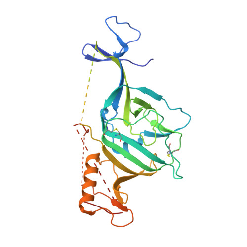 | |
UniProt | |||||
Find proteins for Q05320 (Zaire ebolavirus (strain Mayinga-76)) Explore Q05320 Go to UniProtKB: Q05320 | |||||
Entity Groups | |||||
| Sequence Clusters | 30% Identity50% Identity70% Identity90% Identity95% Identity100% Identity | ||||
| UniProt Group | Q05320 | ||||
Sequence AnnotationsExpand | |||||
| |||||
Find similar proteins by:
(by identity cutoff) | 3D Structure
Entity ID: 2 | |||||
|---|---|---|---|---|---|
| Molecule | Chains | Sequence Length | Organism | Details | Image |
| GP2 | 168 | Ebola virus - Mayinga, Zaire, 1976 | Mutation(s): 1 Gene Names: GP | 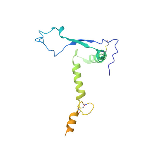 | |
UniProt | |||||
Find proteins for Q05320 (Zaire ebolavirus (strain Mayinga-76)) Explore Q05320 Go to UniProtKB: Q05320 | |||||
Entity Groups | |||||
| Sequence Clusters | 30% Identity50% Identity70% Identity90% Identity95% Identity100% Identity | ||||
| UniProt Group | Q05320 | ||||
Sequence AnnotationsExpand | |||||
| |||||
Oligosaccharides
Entity ID: 3 | |||||
|---|---|---|---|---|---|
| Molecule | Chains | Length | 2D Diagram | Glycosylation | 3D Interactions |
| alpha-D-mannopyranose-(1-6)-beta-D-mannopyranose-(1-4)-2-acetamido-2-deoxy-beta-D-glucopyranose-(1-4)-2-acetamido-2-deoxy-beta-D-glucopyranose | C | 4 |  | N-Glycosylation | |
Glycosylation Resources | |||||
GlyTouCan: G22573RC GlyCosmos: G22573RC GlyGen: G22573RC | |||||
Small Molecules
| Ligands 2 Unique | |||||
|---|---|---|---|---|---|
| ID | Chains | Name / Formula / InChI Key | 2D Diagram | 3D Interactions | |
| YPS Query on YPS | H [auth A] | (1R,3S,5R,7R)-N-[(1r,4R)-4-aminocyclohexyl]-3-(ethoxymethyl)-5-phenyladamantane-1-carboxamide C26 H38 N2 O2 QUVAMRWCRBWKIJ-OKWRYAMFSA-N |  | ||
| NAG Query on NAG | D [auth A], E [auth A], F [auth A], G [auth A] | 2-acetamido-2-deoxy-beta-D-glucopyranose C8 H15 N O6 OVRNDRQMDRJTHS-FMDGEEDCSA-N |  | ||
Experimental Data & Validation
Experimental Data
- Method: X-RAY DIFFRACTION
- Resolution: 2.70 Å
- R-Value Free: 0.225
- R-Value Work: 0.189
- R-Value Observed: 0.192
- Space Group: H 3 2
- Diffraction Data: https://doi.org/10.18430/M37M2D
Unit Cell:
| Length ( Å ) | Angle ( ˚ ) |
|---|---|
| a = 115.11 | α = 90 |
| b = 115.11 | β = 90 |
| c = 309.02 | γ = 120 |
| Software Name | Purpose |
|---|---|
| PHENIX | refinement |
| XDS | data reduction |
| XSCALE | data scaling |
| PDB_EXTRACT | data extraction |
| PHASER | phasing |
| PHENIX | model building |
| Coot | model building |
Entry History
Deposition Data
- Released Date: 2021-11-24 Deposition Author(s): Seattle Structural Genomics Center for Infectious Disease (SSGCID)
Revision History (Full details and data files)
- Version 1.0: 2021-11-24
Type: Initial release - Version 1.1: 2023-10-18
Changes: Data collection, Refinement description
