7MYY

Crystal Structure of HIV-1 PRS17 with GRL-142


Experimental Data Snapshot

  • Method: X-RAY DIFFRACTION
  • Resolution: 1.50 Å
  • R-Value Free: 0.248 
  • R-Value Work: 0.202 
  • R-Value Observed: 0.204 

wwPDB Validation   3D Report Full Report


Ligand Structure Quality Assessment 


This is version 1.1 of the entry. See complete history


Literature

Novel HIV PR inhibitors with C4-substituted bis-THF and bis-fluoro-benzyl target the two active site mutations of highly drug resistant mutant PR S17 .

Agniswamy, J.Kneller, D.W.Ghosh, A.K.Weber, I.T.

(2021) Biochem Biophys Res Commun 566: 30-35

  • DOI: https://doi.org/10.1016/j.bbrc.2021.05.094
  • Primary Citation of Related Structures:  
    7MYP, 7MYY

  • PubMed Abstract: 

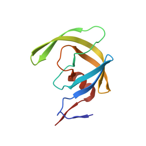
    The emergence of multidrug resistant (MDR) HIV strains severely reduces the effectiveness of antiretroviral therapy. Clinical inhibitor darunavir (1) has picomolar binding affinity for HIV-1 protease (PR), however, drug resistant variants like PR S17 show poor inhibition by 1, despite the presence of only two mutated residues in the inhibitor-binding site. Antiviral inhibitors that target MDR proteases like PR S17 would be valuable as therapeutic agents. Inhibitors 2 and 3 derived from 1 through substitutions at P1, P2 and P2' positions exhibit 3.4- to 500-fold better inhibition than clinical inhibitors for PR S17 with the exception of amprenavir. Crystal structures of PR S17 /2 and PR S17 /3 reveal how these inhibitors target the two active site mutations of PR S17 . The substituted methoxy P2 group of 2 forms new interactions with G48V mutation, while the modified bis-fluoro-benzyl P1 group of 3 forms a halogen interaction with V82S mutation, contributing to improved inhibition of PR S17 .


  • Organizational Affiliation

    Department of Biology, Georgia State University, Atlanta, GA, 30303, USA.


Macromolecules
Find similar proteins by:  (by identity cutoff)  |  3D Structure
Entity ID: 1
MoleculeChains Sequence LengthOrganismDetailsImage
Protease
A, B
99Human immunodeficiency virus 1Mutation(s): 0 
Gene Names: pol
UniProt
Find proteins for I7BFC3 (Human immunodeficiency virus 1)
Explore I7BFC3 
Go to UniProtKB:  I7BFC3
Entity Groups  
Sequence Clusters30% Identity50% Identity70% Identity90% Identity95% Identity100% Identity
UniProt GroupI7BFC3
Sequence Annotations
Expand
  • Reference Sequence
Small Molecules
Ligands 3 Unique
IDChains Name / Formula / InChI Key2D Diagram3D Interactions
7OA (Subject of Investigation/LOI)
Query on 7OA

Download Ideal Coordinates CCD File 
C [auth A](3S,3aR,5R,7aS,8S)-hexahydro-4H-3,5-methanofuro[2,3-b]pyran-8-yl [(2S,3R)-4-[{[2-(cyclopropylamino)-1,3-benzothiazol-6-yl]sulfonyl}(2-methylpropyl)amino]-1-(3,5-difluorophenyl)-3-hydroxybutan-2-yl]carbamate
C33 H40 F2 N4 O7 S2
ARWGTMGGJGADTI-VFGYXJDYSA-N
GOL
Query on GOL

Download Ideal Coordinates CCD File 
D [auth A],
F [auth B]
GLYCEROL
C3 H8 O3
PEDCQBHIVMGVHV-UHFFFAOYSA-N
CL
Query on CL

Download Ideal Coordinates CCD File 
E [auth A]CHLORIDE ION
Cl
VEXZGXHMUGYJMC-UHFFFAOYSA-M
Experimental Data & Validation

Experimental Data

  • Method: X-RAY DIFFRACTION
  • Resolution: 1.50 Å
  • R-Value Free: 0.248 
  • R-Value Work: 0.202 
  • R-Value Observed: 0.204 
  • Space Group: P 61
Unit Cell:
Length ( Å )Angle ( ˚ )
a = 62.893α = 90
b = 62.893β = 90
c = 83.114γ = 120
Software Package:
Software NamePurpose
REFMACrefinement
PDB_EXTRACTdata extraction
HKL-2000data reduction
HKL-2000data scaling
PHASERphasing

Structure Validation

View Full Validation Report


Ligand Structure Quality Assessment 


Entry History & Funding Information

Deposition Data


Funding OrganizationLocationGrant Number
National Institutes of Health/National Cancer Institute (NIH/NCI)United StatesAI150461

Revision History  (Full details and data files)

  • Version 1.0: 2021-07-28
    Type: Initial release
  • Version 1.1: 2023-10-18
    Changes: Data collection, Database references, Refinement description