8A2Y

Cryo-EM structure of F-actin in the Ca2+-ADP-Pi nucleotide state.


Experimental Data Snapshot

  • Method: ELECTRON MICROSCOPY
  • Resolution: 2.15 Å
  • Aggregation State: FILAMENT 
  • Reconstruction Method: SINGLE PARTICLE 

wwPDB Validation   3D Report Full Report


This is version 1.2 of the entry. See complete history


Literature

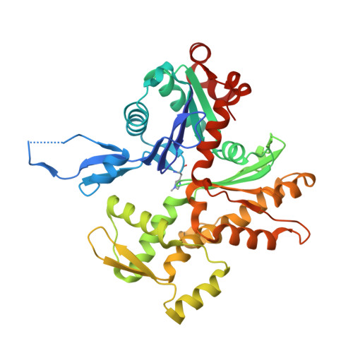
Structural basis of actin filament assembly and aging.

Oosterheert, W.Klink, B.U.Belyy, A.Pospich, S.Raunser, S.

(2022) Nature 611: 374-379

  • DOI: https://doi.org/10.1038/s41586-022-05241-8
  • Primary Citation of Related Structures:  
    8A2R, 8A2S, 8A2T, 8A2U, 8A2Y, 8A2Z

  • PubMed Abstract: 

    The dynamic turnover of actin filaments (F-actin) controls cellular motility in eukaryotes and is coupled to changes in the F-actin nucleotide state 1-3 . It remains unclear how F-actin hydrolyses ATP and subsequently undergoes subtle conformational rearrangements that ultimately lead to filament depolymerization by actin-binding proteins. Here we present cryo-electron microscopy structures of F-actin in all nucleotide states, polymerized in the presence of Mg 2+ or Ca 2+ at approximately 2.2 Å resolution. The structures show that actin polymerization induces the relocation of water molecules in the nucleotide-binding pocket, activating one of them for the nucleophilic attack of ATP. Unexpectedly, the back door for the subsequent release of inorganic phosphate (P i ) is closed in all structures, indicating that P i release occurs transiently. The small changes in the nucleotide-binding pocket after ATP hydrolysis and P i release are sensed by a key amino acid, amplified and transmitted to the filament periphery. Furthermore, differences in the positions of water molecules in the nucleotide-binding pocket explain why Ca 2+ -actin shows slower polymerization rates than Mg 2+ -actin. Our work elucidates the solvent-driven rearrangements that govern actin filament assembly and aging and lays the foundation for the rational design of drugs and small molecules for imaging and therapeutic applications.


  • Organizational Affiliation

    Department of Structural Biochemistry, Max Planck Institute of Molecular Physiology, Dortmund, Germany.


Macromolecules
Find similar proteins by:  (by identity cutoff)  |  3D Structure
Entity ID: 1
MoleculeChains Sequence LengthOrganismDetailsImage
Actin, alpha skeletal muscleA [auth C],
B [auth A],
C [auth B],
D,
E
375Oryctolagus cuniculusMutation(s): 0 
UniProt
Find proteins for P68135 (Oryctolagus cuniculus)
Explore P68135 
Go to UniProtKB:  P68135
Entity Groups  
Sequence Clusters30% Identity50% Identity70% Identity90% Identity95% Identity100% Identity
UniProt GroupP68135
Sequence Annotations
Expand
  • Reference Sequence
Small Molecules
Ligands 3 Unique
IDChains Name / Formula / InChI Key2D Diagram3D Interactions
ADP (Subject of Investigation/LOI)
Query on ADP

Download Ideal Coordinates CCD File 
F [auth C],
I [auth A],
L [auth B],
O [auth D],
R [auth E]
ADENOSINE-5'-DIPHOSPHATE
C10 H15 N5 O10 P2
XTWYTFMLZFPYCI-KQYNXXCUSA-N
PO4 (Subject of Investigation/LOI)
Query on PO4

Download Ideal Coordinates CCD File 
H [auth C],
K [auth A],
N [auth B],
Q [auth D],
T [auth E]
PHOSPHATE ION
O4 P
NBIIXXVUZAFLBC-UHFFFAOYSA-K
CA (Subject of Investigation/LOI)
Query on CA

Download Ideal Coordinates CCD File 
G [auth C],
J [auth A],
M [auth B],
P [auth D],
S [auth E]
CALCIUM ION
Ca
BHPQYMZQTOCNFJ-UHFFFAOYSA-N
Modified Residues  1 Unique
IDChains TypeFormula2D DiagramParent
HIC
Query on HIC
A [auth C],
B [auth A],
C [auth B],
D,
E
L-PEPTIDE LINKINGC7 H11 N3 O2HIS
Experimental Data & Validation

Experimental Data

  • Method: ELECTRON MICROSCOPY
  • Resolution: 2.15 Å
  • Aggregation State: FILAMENT 
  • Reconstruction Method: SINGLE PARTICLE 
EM Software:
TaskSoftware PackageVersion
MODEL REFINEMENTPHENIXv1.20.1-4487
RECONSTRUCTIONRELION3.1.0

Structure Validation

View Full Validation Report


Entry History & Funding Information

Deposition Data


Funding OrganizationLocationGrant Number
European Research Council (ERC)European Union856118
Alexander von Humboldt FoundationGermany--
European Molecular Biology Organization (EMBO)European Union--
Max Planck SocietyGermany--

Revision History  (Full details and data files)

  • Version 1.0: 2022-08-10
    Type: Initial release
  • Version 1.1: 2022-11-16
    Changes: Database references
  • Version 1.2: 2022-11-23
    Changes: Database references