5BMH

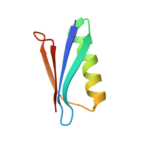
Nitroxide Spin Labels in Protein GB1: T44 Mutant, Crystal Form B


Experimental Data Snapshot

  • Method: X-RAY DIFFRACTION
  • Resolution: 1.60 Å
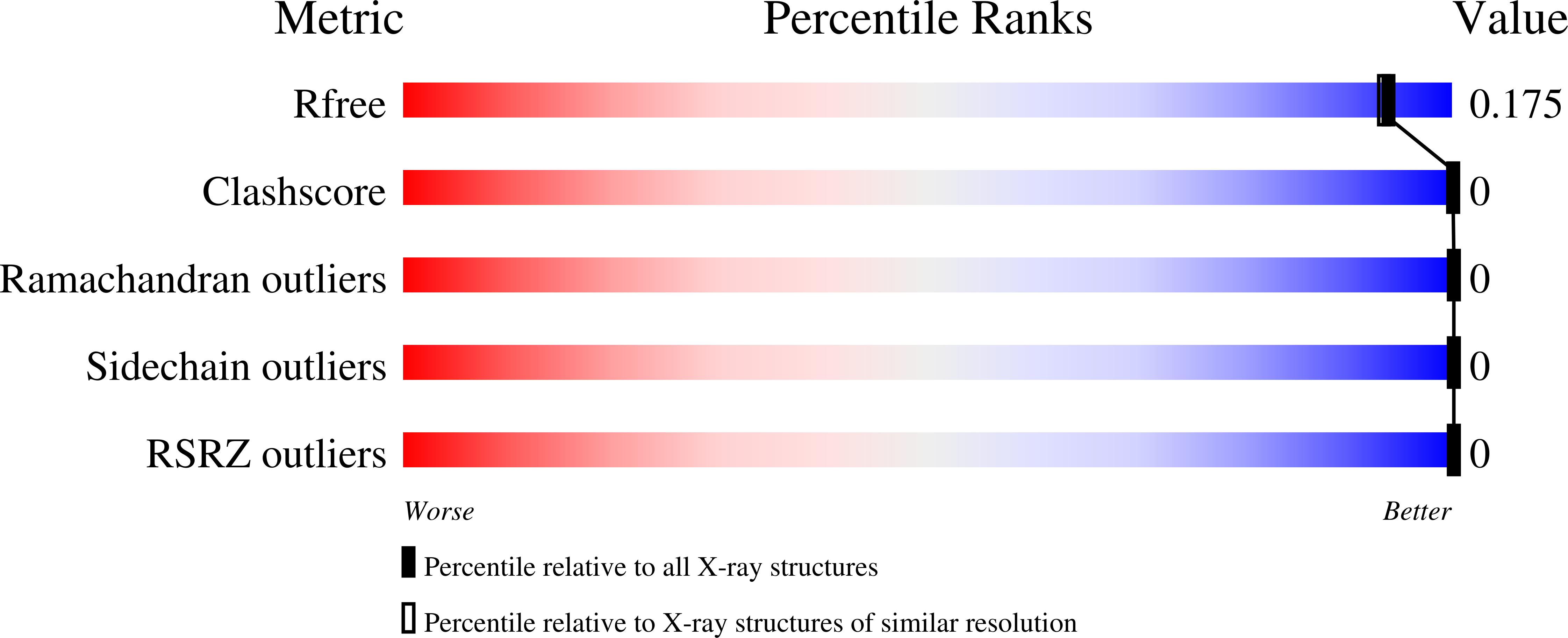
  • R-Value Free: 0.175 
  • R-Value Work: 0.155 
  • R-Value Observed: 0.156 

Starting Model: experimental
View more details

wwPDB Validation   3D Report Full Report


Ligand Structure Quality Assessment 


This is version 1.6 of the entry. See complete history


Literature

Rotameric preferences of a protein spin label at edge-strand beta-sheet sites.

Cunningham, T.F.Pornsuwan, S.Horne, W.S.Saxena, S.

(2016) Protein Sci 25: 1049-1060

  • DOI: https://doi.org/10.1002/pro.2918
  • Primary Citation of Related Structures:  
    5BMG, 5BMH, 5BMI

  • PubMed Abstract: 

    Protein spin labeling to yield the nitroxide-based R1 side chain is a powerful method to measure protein dynamics and structure by electron spin resonance. However, R1 measurements are complicated by the flexibility of the side chain. While analysis approaches for solvent-exposed α-helical environment have been developed to partially account for flexibility, similar work in β-sheets is lacking. The goal of this study is to provide the first essential steps for understanding the conformational preferences of R1 within edge β-strands using X-ray crystallography and double electron electron resonance (DEER) distance measurements. Crystal structures yielded seven rotamers for a non-hydrogen-bonded site and three rotamers for a hydrogen-bonded site. The observed rotamers indicate contextual differences in R1 conformational preferences compared to other solvent-exposed environments. For the DEER measurements, each strand site was paired with the same α-helical site elsewhere on the protein. The most probable distance observed by DEER is rationalized based on the rotamers observed in the crystal structure. Additionally, the appropriateness of common molecular modeling methods that account for R1 conformational preferences are assessed for the β-sheet environment. These results show that interpretation of R1 behavior in β-sheets is difficult and indicate further development is needed for these computational methods to correctly relate DEER distances to protein structure at edge β-strand sites.


  • Organizational Affiliation

    Department of Chemistry, University of Pittsburgh, 219 Parkman Ave, Pittsburgh, Pennsylvania, 15260.


Macromolecules
Find similar proteins by:  (by identity cutoff)  |  3D Structure
Entity ID: 1
MoleculeChains Sequence LengthOrganismDetailsImage
Immunoglobulin G-binding protein G56Streptococcus sp. 'group GMutation(s): 1 
Gene Names: spg
UniProt
Find proteins for P19909 (Streptococcus sp. group G)
Explore P19909 
Go to UniProtKB:  P19909
Entity Groups  
Sequence Clusters30% Identity50% Identity70% Identity90% Identity95% Identity100% Identity
UniProt GroupP19909
Sequence Annotations
Expand
  • Reference Sequence
Small Molecules
Ligands 1 Unique
IDChains Name / Formula / InChI Key2D Diagram3D Interactions
MTN
Query on MTN

Download Ideal Coordinates CCD File 
B [auth A]S-[(1-oxyl-2,2,5,5-tetramethyl-2,5-dihydro-1H-pyrrol-3-yl)methyl] methanesulfonothioate
C10 H18 N O3 S2
MXZPGYFBZHBAQM-UHFFFAOYSA-N
Experimental Data & Validation

Experimental Data

  • Method: X-RAY DIFFRACTION
  • Resolution: 1.60 Å
  • R-Value Free: 0.175 
  • R-Value Work: 0.155 
  • R-Value Observed: 0.156 
  • Space Group: P 21 21 21
Unit Cell:
Length ( Å )Angle ( ˚ )
a = 25.037α = 90
b = 37.194β = 90
c = 49.217γ = 90
Software Package:
Software NamePurpose
PHENIXrefinement
CrystalCleardata reduction
CrystalCleardata scaling

Structure Validation

View Full Validation Report


Ligand Structure Quality Assessment 


Entry History & Funding Information

Deposition Data


Funding OrganizationLocationGrant Number
National Science Foundation (NSF, United States)United StatesMCB-1157712

Revision History  (Full details and data files)

  • Version 1.0: 2016-04-06
    Type: Initial release
  • Version 1.1: 2016-05-04
    Changes: Database references
  • Version 1.2: 2017-09-13
    Changes: Author supporting evidence, Database references, Derived calculations
  • Version 1.3: 2019-11-27
    Changes: Author supporting evidence
  • Version 1.4: 2023-09-27
    Changes: Data collection, Database references, Refinement description
  • Version 1.5: 2024-05-01
    Changes: Advisory, Derived calculations
  • Version 1.6: 2024-11-06
    Changes: Structure summary