7MX3

Crystal structure of human RIPK3 complexed with GSK'843


Experimental Data Snapshot

  • Method: X-RAY DIFFRACTION
  • Resolution: 3.23 Å
  • R-Value Free: 0.285 
  • R-Value Work: 0.239 
  • R-Value Observed: 0.244 

wwPDB Validation   3D Report Full Report


Ligand Structure Quality Assessment 


This is version 1.2 of the entry. See complete history


Literature

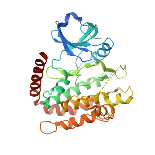
Human RIPK3 maintains MLKL in an inactive conformation prior to cell death by necroptosis.

Meng, Y.Davies, K.A.Fitzgibbon, C.Young, S.N.Garnish, S.E.Horne, C.R.Luo, C.Garnier, J.M.Liang, L.Y.Cowan, A.D.Samson, A.L.Lessene, G.Sandow, J.J.Czabotar, P.E.Murphy, J.M.

(2021) Nat Commun 12: 6783-6783

  • DOI: https://doi.org/10.1038/s41467-021-27032-x
  • Primary Citation of Related Structures:  
    7MON, 7MX3

  • PubMed Abstract: 

    The ancestral origins of the lytic cell death mode, necroptosis, lie in host defense. However, the dysregulation of necroptosis in inflammatory diseases has led to widespread interest in targeting the pathway therapeutically. This mode of cell death is executed by the terminal effector, the MLKL pseudokinase, which is licensed to kill following phosphorylation by its upstream regulator, RIPK3 kinase. The precise molecular details underlying MLKL activation are still emerging and, intriguingly, appear to mechanistically-diverge between species. Here, we report the structure of the human RIPK3 kinase domain alone and in complex with the MLKL pseudokinase. These structures reveal how human RIPK3 structurally differs from its mouse counterpart, and how human RIPK3 maintains MLKL in an inactive conformation prior to induction of necroptosis. Residues within the RIPK3:MLKL C-lobe interface are crucial to complex assembly and necroptotic signaling in human cells, thereby rationalizing the strict species specificity governing RIPK3 activation of MLKL.


  • Organizational Affiliation

    Walter and Eliza Hall Institute of Medical Research, 1G Royal Parade, Parkville, VIC, 3052, Australia.


Macromolecules
Find similar proteins by:  (by identity cutoff)  |  3D Structure
Entity ID: 1
MoleculeChains Sequence LengthOrganismDetailsImage
Receptor-interacting serine/threonine-protein kinase 3
A, B, C, D
319Homo sapiensMutation(s): 2 
Gene Names: RIPK3RIP3
EC: 2.7.11.1
UniProt & NIH Common Fund Data Resources
Find proteins for Q9Y572 (Homo sapiens)
Explore Q9Y572 
Go to UniProtKB:  Q9Y572
PHAROS:  Q9Y572
GTEx:  ENSG00000129465 
Entity Groups  
Sequence Clusters30% Identity50% Identity70% Identity90% Identity95% Identity100% Identity
UniProt GroupQ9Y572
Sequence Annotations
Expand
  • Reference Sequence
Experimental Data & Validation

Experimental Data

  • Method: X-RAY DIFFRACTION
  • Resolution: 3.23 Å
  • R-Value Free: 0.285 
  • R-Value Work: 0.239 
  • R-Value Observed: 0.244 
  • Space Group: P 21 21 21
Unit Cell:
Length ( Å )Angle ( ˚ )
a = 104.823α = 90
b = 104.823β = 90
c = 126.677γ = 90
Software Package:
Software NamePurpose
PHENIXrefinement
Aimlessdata scaling
PDB_EXTRACTdata extraction
DIALSdata reduction
autoPROCdata processing
PHASERphasing

Structure Validation

View Full Validation Report


Ligand Structure Quality Assessment 


Entry History & Funding Information

Deposition Data


Funding OrganizationLocationGrant Number
National Health and Medical Research Council (NHMRC, Australia)Australia1172929

Revision History  (Full details and data files)

  • Version 1.0: 2021-11-24
    Type: Initial release
  • Version 1.1: 2021-12-08
    Changes: Database references
  • Version 1.2: 2024-04-03
    Changes: Data collection, Refinement description