7RLE

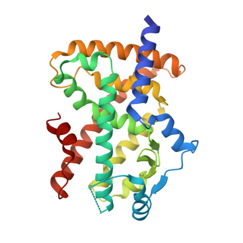
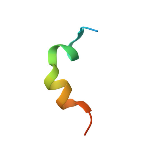
Crystal structure of PPAR gamma in complex with CREB-binding protein and agonist GW1929

  • Classification: DNA BINDING PROTEIN
  • Organism(s): Homo sapiens
  • Expression System: Escherichia coli
  • Mutation(s): No 

  • Deposited: 2021-07-23 Released: 2022-08-03 
  • Deposition Author(s): Nemetchek, M.D., Sprang, S.R., Hughes, T.S.
  • Funding Organization(s): National Institutes of Health/National Institute of General Medical Sciences (NIH/NIGMS), National Institutes of Health/National Institute of Diabetes and Digestive and Kidney Disease (NIH/NIDDK)

Experimental Data Snapshot

  • Method: X-RAY DIFFRACTION
  • Resolution: 2.50 Å
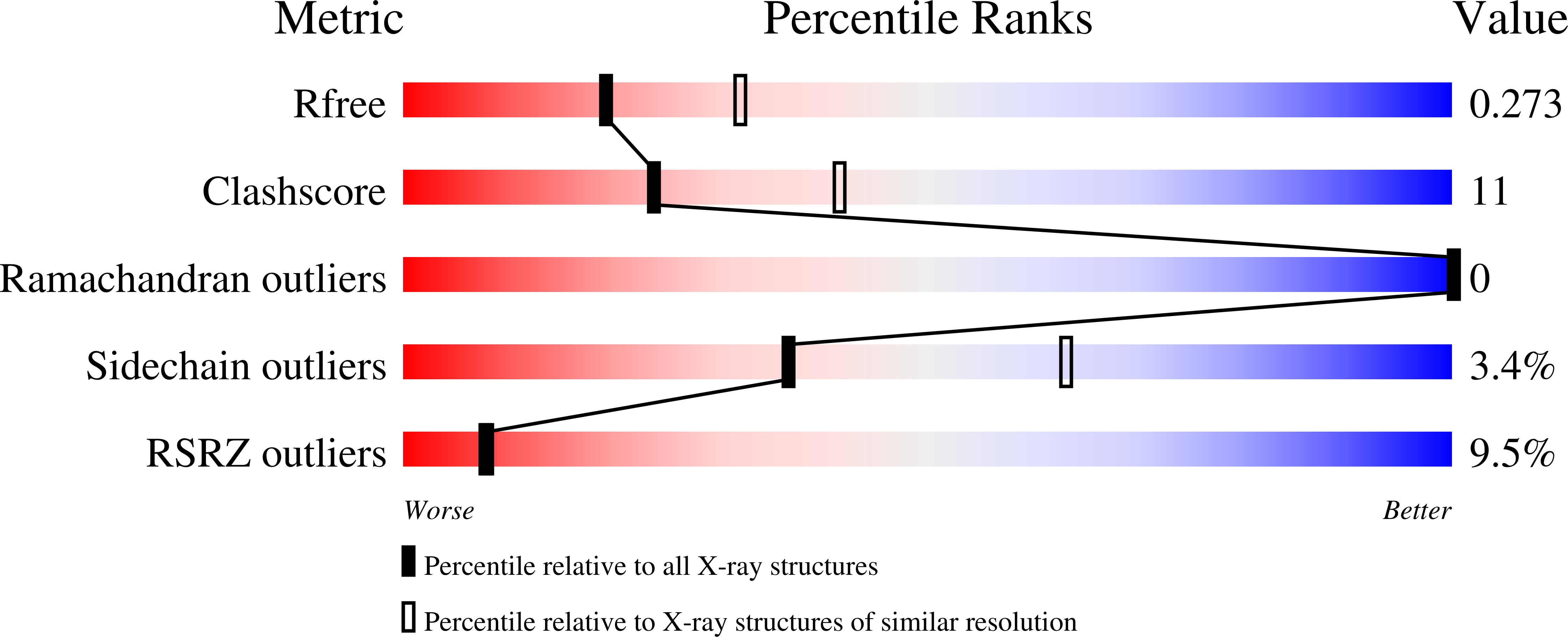
  • R-Value Free: 0.275 
  • R-Value Work: 0.232 
  • R-Value Observed: 0.235 

wwPDB Validation   3D Report Full Report


Ligand Structure Quality Assessment 


This is version 1.2 of the entry. See complete history


Literature

A structural mechanism of nuclear receptor biased agonism.

Nemetchek, M.D.Chrisman, I.M.Rayl, M.L.Voss, A.H.Hughes, T.S.

(2022) Proc Natl Acad Sci U S A 119: e2215333119-e2215333119

  • DOI: https://doi.org/10.1073/pnas.2215333119
  • Primary Citation of Related Structures:  
    6D94, 7RLE

  • PubMed Abstract: 

    Efforts to decrease the adverse effects of nuclear receptor (NR) drugs have yielded experimental agonists that produce better outcomes in mice. Some of these agonists have been shown to cause different, not just less intense, on-target transcriptomic effects; however, a structural explanation for such agonist-specific effects remains unknown. Here, we show that partial agonists of the NR peroxisome proliferator-associated receptor γ (PPARγ), which induce better outcomes in mice compared to clinically utilized type II diabetes PPARγ-binding drugs thiazolidinediones (TZDs), also favor a different group of coactivator peptides than the TZDs. We find that PPARγ full agonists can also be biased relative to each other in terms of coactivator peptide binding. We find differences in coactivator-PPARγ bonding between the coactivator subgroups which allow agonists to favor one group of coactivator peptides over another, including differential bonding to a C-terminal residue of helix 4. Analysis of all available NR-coactivator structures indicates that such differential helix 4 bonding persists across other NR-coactivator complexes, providing a general structural mechanism of biased agonism for many NRs. Further work will be necessary to determine if such bias translates into altered coactivator occupancy and physiology in cells.


  • Organizational Affiliation

    Center for Biomolecular Structure and Dynamics, University of Montana, Missoula 59812.


Macromolecules
Find similar proteins by:  (by identity cutoff)  |  3D Structure
Entity ID: 1
MoleculeChains Sequence LengthOrganismDetailsImage
Peroxisome proliferator-activated receptor gamma
A, C
276Homo sapiensMutation(s): 0 
Gene Names: PPARGNR1C3
UniProt & NIH Common Fund Data Resources
Find proteins for P37231 (Homo sapiens)
Explore P37231 
Go to UniProtKB:  P37231
PHAROS:  P37231
GTEx:  ENSG00000132170 
Entity Groups  
Sequence Clusters30% Identity50% Identity70% Identity90% Identity95% Identity100% Identity
UniProt GroupP37231
Sequence Annotations
Expand
  • Reference Sequence

Find similar proteins by:  Sequence   |   3D Structure  

Entity ID: 2
MoleculeChains Sequence LengthOrganismDetailsImage
CREB-binding protein
B, D
24Homo sapiensMutation(s): 0 
EC: 2.3.1.48 (PDB Primary Data), 2.3.1 (PDB Primary Data)
UniProt & NIH Common Fund Data Resources
Find proteins for Q92793 (Homo sapiens)
Explore Q92793 
Go to UniProtKB:  Q92793
PHAROS:  Q92793
GTEx:  ENSG00000005339 
Entity Groups  
Sequence Clusters30% Identity50% Identity70% Identity90% Identity95% Identity100% Identity
UniProt GroupQ92793
Sequence Annotations
Expand
  • Reference Sequence
Small Molecules
Ligands 1 Unique
IDChains Name / Formula / InChI Key2D Diagram3D Interactions
EDK (Subject of Investigation/LOI)
Query on EDK

Download Ideal Coordinates CCD File 
E [auth A],
F [auth C]
(2~{S})-3-[4-[2-[methyl(pyridin-2-yl)amino]ethoxy]phenyl]-2-[[2-(phenylcarbonyl)phenyl]amino]propanoic acid
C30 H29 N3 O4
QTQMRBZOBKYXCG-MHZLTWQESA-N
Experimental Data & Validation

Experimental Data

  • Method: X-RAY DIFFRACTION
  • Resolution: 2.50 Å
  • R-Value Free: 0.275 
  • R-Value Work: 0.232 
  • R-Value Observed: 0.235 
  • Space Group: P 21 21 21
Unit Cell:
Length ( Å )Angle ( ˚ )
a = 59.89α = 90
b = 99.422β = 90
c = 109.179γ = 90
Software Package:
Software NamePurpose
PHENIXrefinement
HKL-3000data reduction
HKL-3000data scaling
PHASERphasing

Structure Validation

View Full Validation Report


Ligand Structure Quality Assessment 


Entry History & Funding Information

Deposition Data


Funding OrganizationLocationGrant Number
National Institutes of Health/National Institute of General Medical Sciences (NIH/NIGMS)United StatesP20GM103546
National Institutes of Health/National Institute of Diabetes and Digestive and Kidney Disease (NIH/NIDDK)United StatesR00DK103116

Revision History  (Full details and data files)

  • Version 1.0: 2022-08-03
    Type: Initial release
  • Version 1.1: 2023-03-01
    Changes: Database references
  • Version 1.2: 2023-10-25
    Changes: Data collection, Refinement description