7SVV

TnsBctd-TnsC complex


Experimental Data Snapshot

  • Method: ELECTRON MICROSCOPY
  • Resolution: 3.54 Å
  • Aggregation State: PARTICLE 
  • Reconstruction Method: HELICAL 

wwPDB Validation   3D Report Full Report


This is version 1.2 of the entry. See complete history


Literature

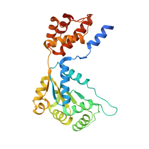
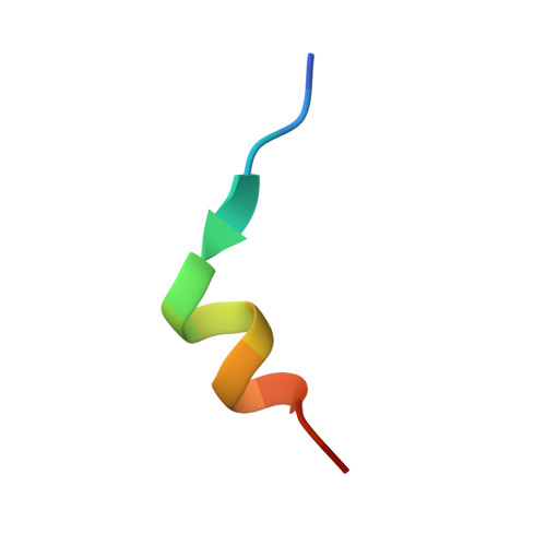
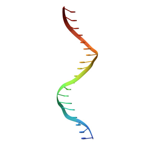
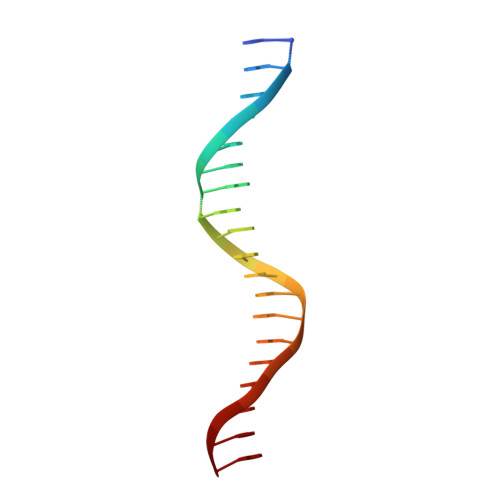
Mechanistic details of CRISPR-associated transposon recruitment and integration revealed by cryo-EM.

Park, J.U.Tsai, A.W.Chen, T.H.Peters, J.E.Kellogg, E.H.

(2022) Proc Natl Acad Sci U S A 119: e2202590119-e2202590119

  • DOI: https://doi.org/10.1073/pnas.2202590119
  • Primary Citation of Related Structures:  
    7SVV, 7SVW

  • PubMed Abstract: 

    CRISPR-associated transposons (CASTs) are Tn7-like elements that are capable of RNA-guided DNA integration. Although structural data are known for nearly all core transposition components, the transposase component, TnsB, remains uncharacterized. Using cryo-electron microscopy (cryo-EM) structure determination, we reveal the conformation of TnsB during transposon integration for the type V-K CAST system from Scytonema hofmanni (ShCAST). Our structure of TnsB is a tetramer, revealing strong mechanistic relationships with the overall architecture of RNaseH transposases/integrases in general, and in particular the MuA transposase from bacteriophage Mu. However, key structural differences in the C-terminal domains indicate that TnsB's tetrameric architecture is stabilized by a different set of protein-protein interactions compared with MuA. We describe the base-specific interactions along the TnsB binding site, which explain how different CAST elements can function on cognate mobile elements independent of one another. We observe that melting of the 5' nontransferred strand of the transposon end is a structural feature stabilized by TnsB and furthermore is crucial for donor-DNA integration. Although not observed in the TnsB strand-transfer complex, the C-terminal end of TnsB serves a crucial role in transposase recruitment to the target site. The C-terminal end of TnsB adopts a short, structured 15-residue "hook" that decorates TnsC filaments. Unlike full-length TnsB, C-terminal fragments do not appear to stimulate filament disassembly using two different assays, suggesting that additional interactions between TnsB and TnsC are required for redistributing TnsC to appropriate targets. The structural information presented here will help guide future work in modifying these important systems as programmable gene integration tools.


  • Organizational Affiliation

    Department of Molecular Biology and Genetics, Cornell University, Ithaca, NY 14853.


Macromolecules

Find similar proteins by:  (by identity cutoff)  |  3D Structure
Entity ID: 3
MoleculeChains Sequence LengthOrganismDetailsImage
TnsC filament276[Scytonema hofmanni] UTEX 2349Mutation(s): 0 
UniProt
Find proteins for A0A8J0PCL3 (Scytonema hofmannii)
Explore A0A8J0PCL3 
Go to UniProtKB:  A0A8J0PCL3
Entity Groups  
Sequence Clusters30% Identity50% Identity70% Identity90% Identity95% Identity100% Identity
UniProt GroupA0A8J0PCL3
Sequence Annotations
Expand
  • Reference Sequence

Find similar proteins by:  Sequence   |   3D Structure  

Entity ID: 4
MoleculeChains Sequence LengthOrganismDetailsImage
TnsBctd15[Scytonema hofmanni] UTEX 2349Mutation(s): 0 
Entity Groups  
Sequence Clusters30% Identity50% Identity70% Identity90% Identity95% Identity100% Identity
Sequence Annotations
Expand
  • Reference Sequence

Find similar nucleic acids by:  Sequence   |   3D Structure  

Entity ID: 1
MoleculeChains LengthOrganismImage
DNA (5'-D(P*TP*TP*TP*TP*TP*TP*TP*TP*TP*TP*TP*TP*TP*TP*TP*TP*TP*TP*T)-3')A [auth 1]19synthetic construct
Sequence Annotations
Expand
  • Reference Sequence

Find similar nucleic acids by:  Sequence   |   3D Structure  

Entity ID: 2
MoleculeChains LengthOrganismImage
DNA (5'-D(P*AP*AP*AP*AP*AP*AP*AP*AP*AP*AP*AP*AP*AP*AP*AP*AP*AP*AP*A)-3')B [auth 2]22synthetic construct
Sequence Annotations
Expand
  • Reference Sequence
Small Molecules
Ligands 2 Unique
IDChains Name / Formula / InChI Key2D Diagram3D Interactions
ANP
Query on ANP

Download Ideal Coordinates CCD File 
AA [auth C]
CA [auth D]
EA [auth E]
GA [auth F]
IA [auth G]
AA [auth C],
CA [auth D],
EA [auth E],
GA [auth F],
IA [auth G],
KA [auth H],
MA [auth I],
OA [auth J],
W [auth A],
Y [auth B]
PHOSPHOAMINOPHOSPHONIC ACID-ADENYLATE ESTER
C10 H17 N6 O12 P3
PVKSNHVPLWYQGJ-KQYNXXCUSA-N
MG
Query on MG

Download Ideal Coordinates CCD File 
BA [auth C]
DA [auth D]
FA [auth E]
HA [auth F]
JA [auth G]
BA [auth C],
DA [auth D],
FA [auth E],
HA [auth F],
JA [auth G],
LA [auth H],
NA [auth I],
PA [auth J],
X [auth A],
Z [auth B]
MAGNESIUM ION
Mg
JLVVSXFLKOJNIY-UHFFFAOYSA-N
Experimental Data & Validation

Experimental Data

  • Method: ELECTRON MICROSCOPY
  • Resolution: 3.54 Å
  • Aggregation State: PARTICLE 
  • Reconstruction Method: HELICAL 

Structure Validation

View Full Validation Report


Entry History & Funding Information

Deposition Data


Funding OrganizationLocationGrant Number
National Institutes of Health/National Institute of General Medical Sciences (NIH/NIGMS)United StatesR00-GM124463

Revision History  (Full details and data files)

  • Version 1.0: 2022-08-10
    Type: Initial release
  • Version 1.1: 2022-08-17
    Changes: Database references
  • Version 1.2: 2024-02-28
    Changes: Data collection, Refinement description