


New Features
COVID-19/SARS-CoV-2 Resources
03/25
RCSB.org/covid19:
PDB Structures (as of July 24, 2024)
PDB Structures (as of July 24, 2024)
Access all SARS-CoV-2 PDB structures
- New this week
- Main proteases
- Spike proteins and receptor binding domains
- Papain-like proteinases
- Other SARS-CoV-2 structures
- PanDDA analysis: main protease | helicase | NendoU | Nsp3 macrodomain | NSP14
- COVID MOONSHOT
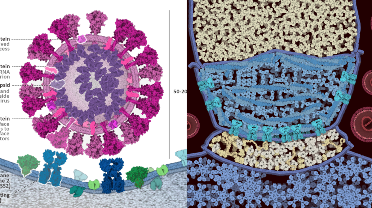
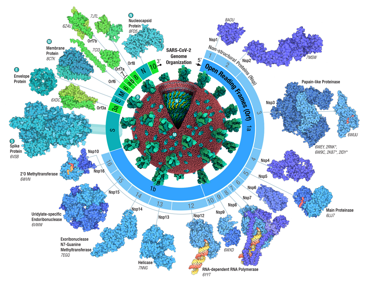 Adapted from Architecture of the SARS-CoV-2 genome and proteome in Evolution of the SARS-CoV-2 proteome in three dimensions (3D) during the first 6 months of the COVID-19 pandemic Proteins: Structure, Function, and Bioinformatics (2022) 90: 1054-1080; doi: 10.1002/prot.26250
Scientific Publications
- Genetic and Structural Analysis of SARS-CoV-2 Spike Protein for Universal Epitope Selection (2022) Molecular Biology and Evolution 39: msac091 doi: 10.1093/molbev/msac091
- Structural models of SARS-CoV-2 Omicron variant in complex with ACE2 receptor or antibodies suggest altered binding interfaces (2021) bioRxiv doi: 10.1101/2021.12.12.472313
- Design and proof of concept for targeted phage-based COVID-19 vaccination strategies with a streamlined cold-free supply chain (2021) PNAS 118: e21057391181 doi: 10.1073/pnas.2105739118
- Structure-enabled Design of mRNA Vaccines (2021) Structure 10.1016/j.str.2021.10.008
- Architecture of the SARS-CoV-2 genome and proteome (2021) PROTEINS doi: 10.1002/prot.26250
PDB-101 Educational Resources and Images (all free for use)
- Educational resources at PDB-101
- Molecule of the Month features: Coronavirus Proteases | SARS-CoV-2 Spike | SARS-CoV-2 RNA-dependent RNA Polymerase | SARS-CoV-2 Spike Variants
- Resources to Fight the COVID-19 Pandemic: Passive Immunization with Convalescent Antibodies | The Search for Drugs to Fight COVID-19 | Dexamethasone and Cytokine Storms | SARS-CoV-2 mRNA Vaccine
- Boot Camp: COVID-19 Evolution and Structural Biology Week-long experience with undergraduates to study sequence and structure changes during the first six months of the pandemic (see also BAMBed (2020) doi: 10.1002/bmb.21428)
- Curriculum Modules: COVID-19 in Molecular Detail
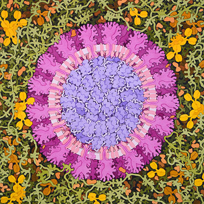
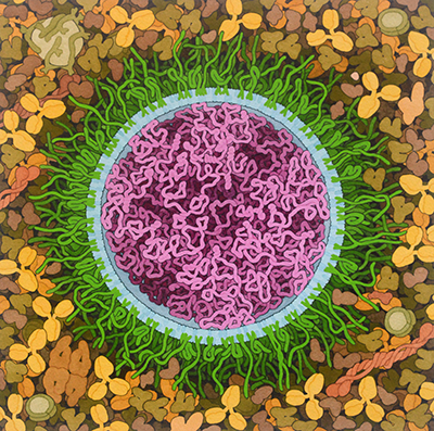 SARS-CoV-2 mRNA Vaccine, 2020
David S. Goodsell, RCSB PDB
doi: 10.2210/rcsb_pdb/goodsell-gallery-027

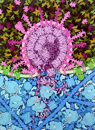

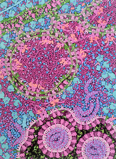
SARS-CoV-2 Fusion, 2020
David S. Goodsell, RCSB PDB doi: 10.2210/rcsb_pdb/goodsell-gallery-026
Respiratory Droplet, 2020
David S. Goodsell, RCSB PDB doi: 10.2210/rcsb_pdb/goodsell-gallery-024
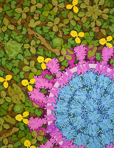
SARS-CoV-2 and Neutralizing Antibodies, 2020
David S. Goodsell, RCSB PDB doi: 10.2210/rcsb_pdb/goodsell-gallery-025
Video: Fighting Coronavirus with Soap
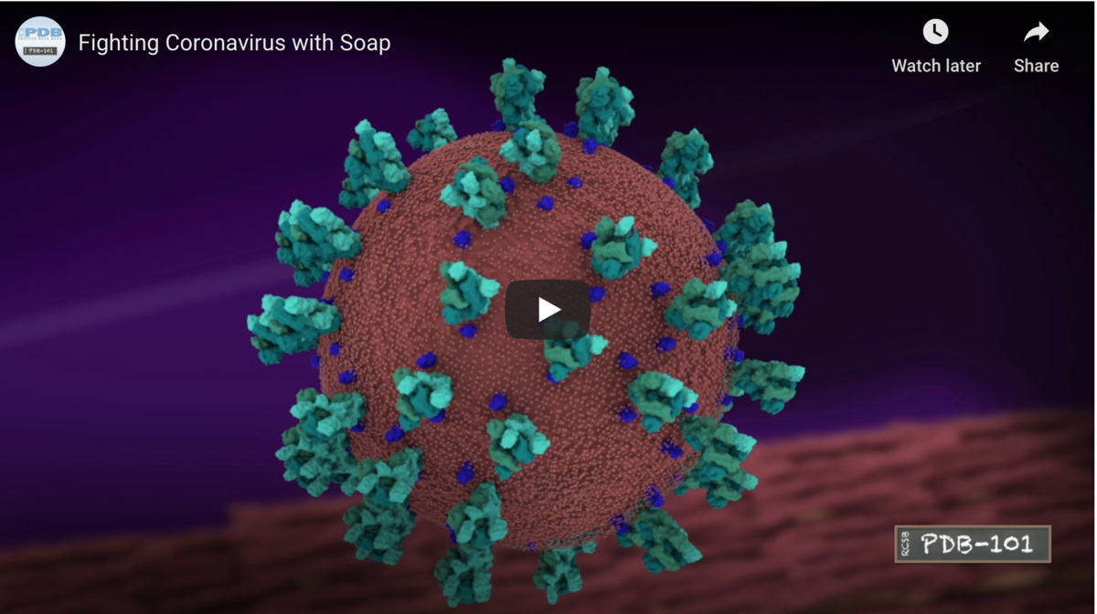 Video: Watch at the molecular level how soap breaks up coronavirus.
 Animated GIF
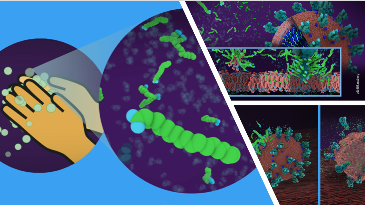 Video still images for download
Images
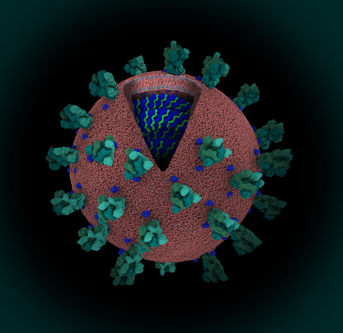
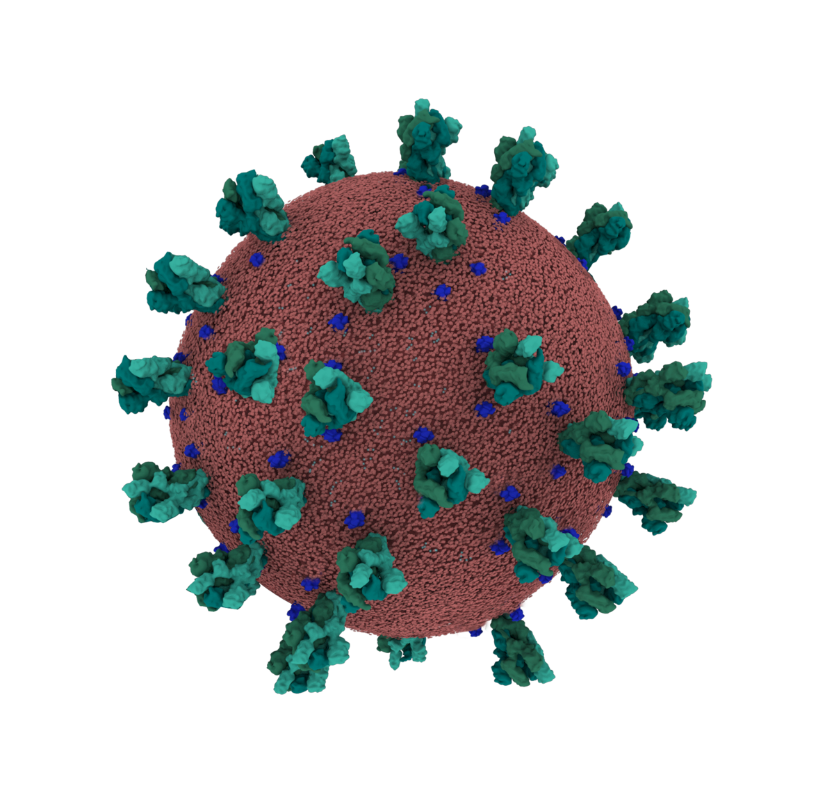 Coronavirus Credit: Maria Voigt/RCSB PDB
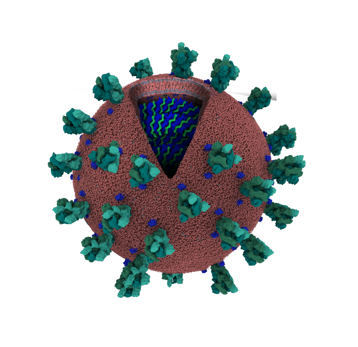

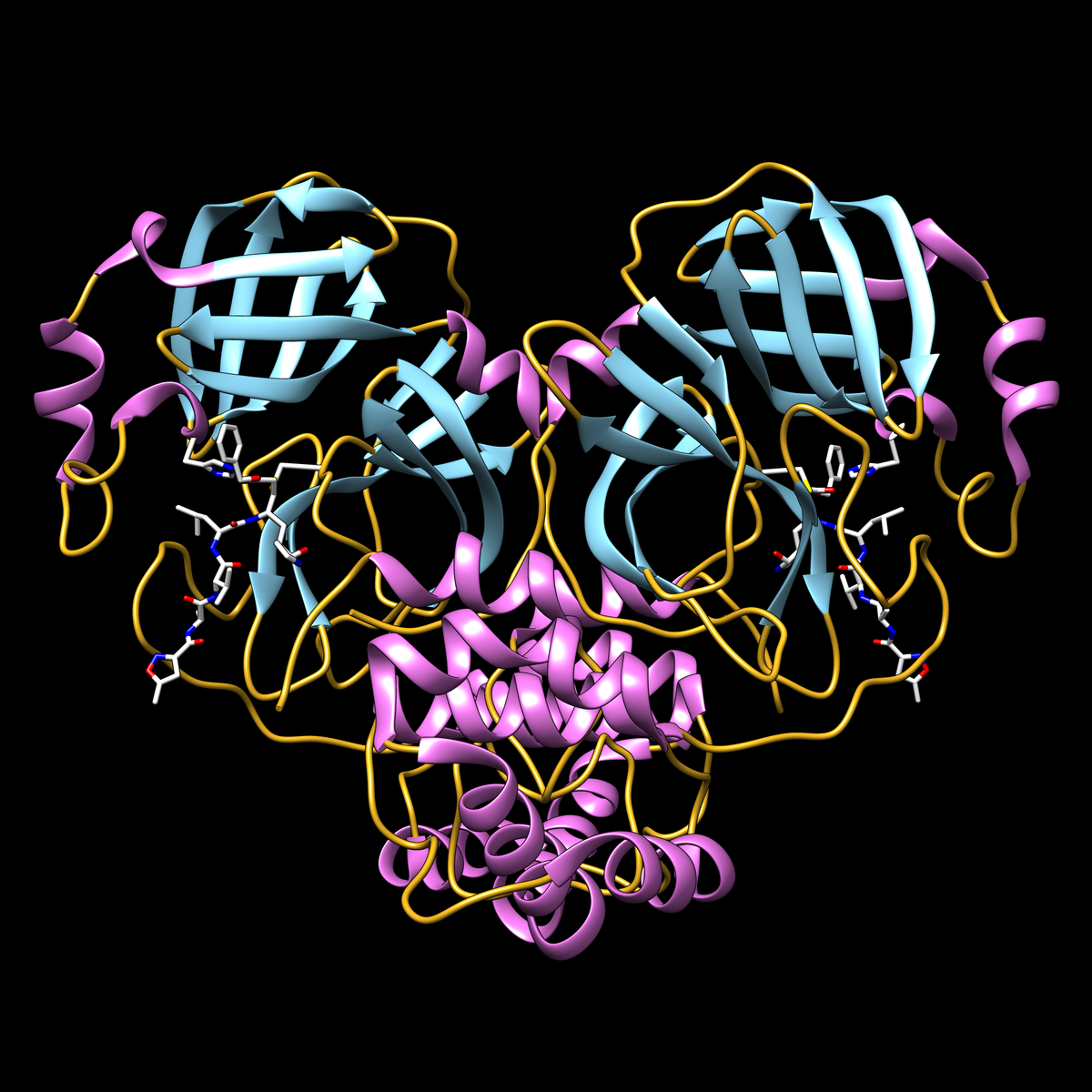 PDB structure 6LU7 of SARS-CoV-2 main protease
Video
Main protease protein with inhibitor N3 (white stick representation) covalently bound to residue cysteine 145 in the protease active site. Display shows secondary structure (helices in magenta, strands in cyan, loops in yellow). Adjacent active site residue histidine 41 is also shown.
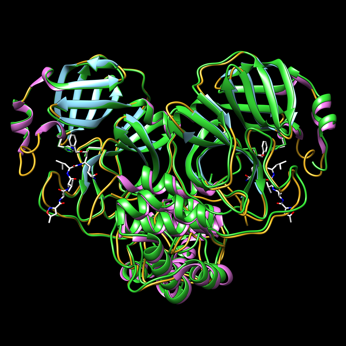 Structural similarity of the SARS-CoV-2 main protease (PDB structure 6lu7) and the SARS coronavirus main protease (PDB structure 1q2w in green)
Video
 PDB structure 6LU7 of SARS-CoV-2 main protease
Video
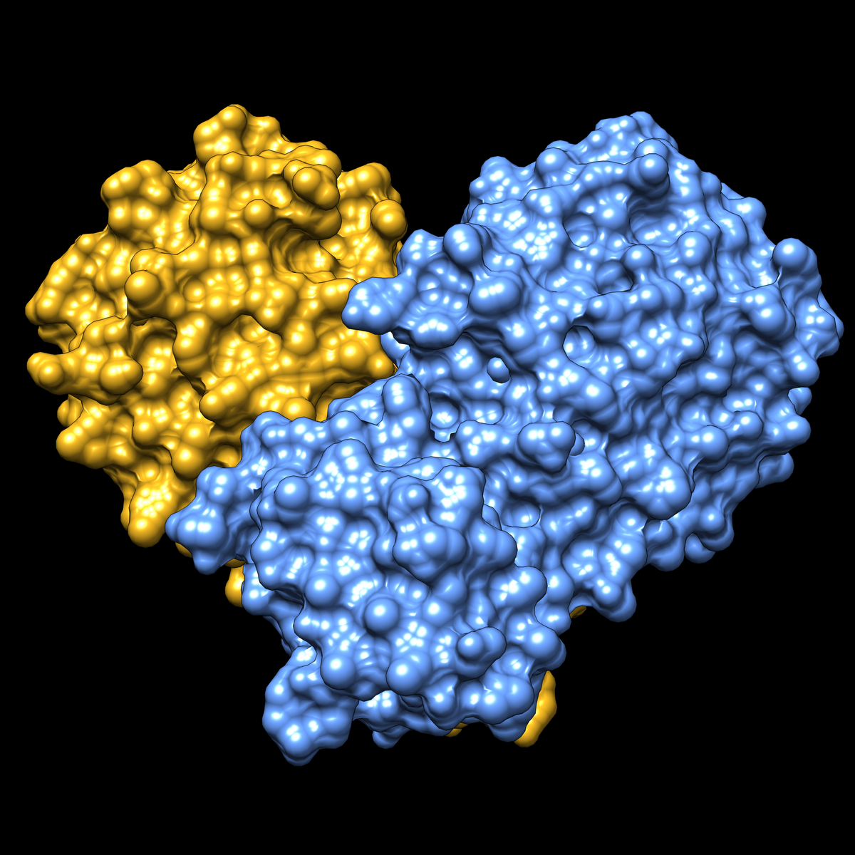
Shown in a surface representation, with the two halves of the dimer in yellow and blue.
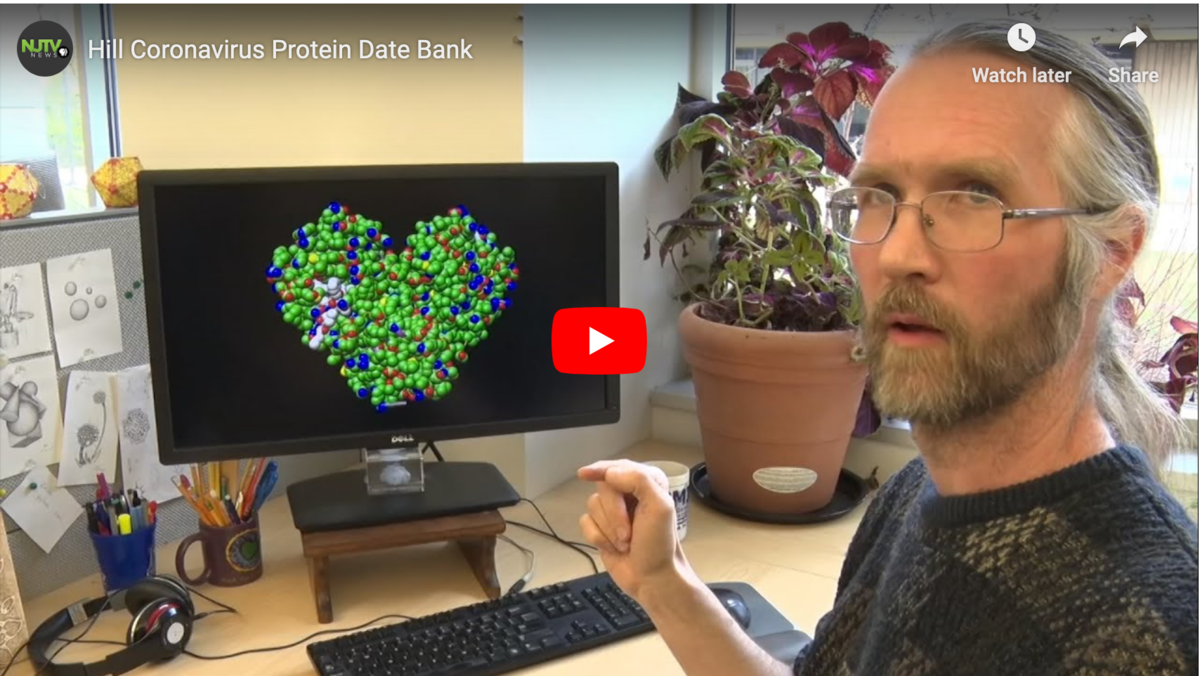 NJTV News talks with Rutgers University researchers Stephen Burley and Brian Hudson at the Protein Data Bank about finding the key to fighting COVID-19.
 Coronavirus main protease virtual meeting background for download;
based on PDB structure 6lu7
Coronavirus main protease virtual meeting background for download;
based on PDB structure 6lu7Other backgrounds are available for download from PDB-101
Background Information and Select Media Mentions
- Structure of the Pandemic by Sarah Kearns (also at Structure (2020) 28: 874-878 doi: 10.1016/j.str.2020.07.007
- PDB structure 6lu7: first high-resolution crystal structure of SARS-CoV-2 main protease (released February 5, 2020). doi: 10.2210/pdb6lu7/pdb
- wwPDB announcement, including summary data for closely-related PDB structures
- How to help the free market fight coronavirus (2020) Nature 580: 167 doi: 10.1038/d41586-020-00888-7
- NJTV News video segment on the PDB and Cracking the Coronavirus Code
- Bad News Wrapped in Protein: Inside the Coronavirus Genome The New York Times April 3
- COVID19 in VR: RCSB Protein Data Bank Biocurator Joins Us Inside Nanome (Video). Biocurator Dr. Gregg Crichlow joined Nanome inside VR to look at two SARS-CoV-2 spike protein structures from the PDB.
Additional Resources
RCSB PDB (citation) is hosted by
RCSB PDB is a member of the
