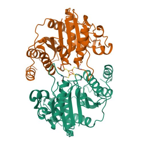
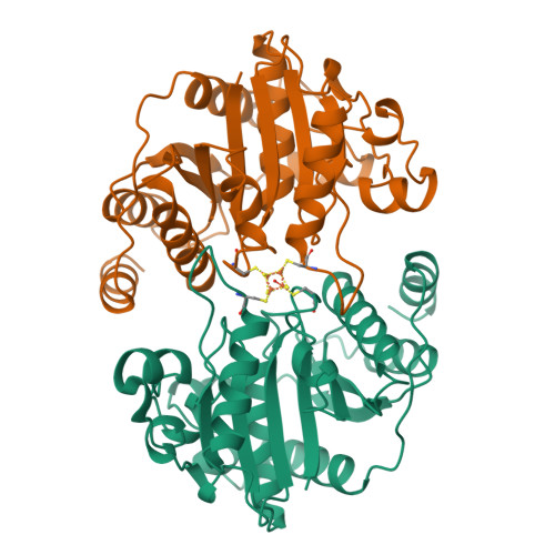
Conformational variability in structures of the nitrogenase iron proteins from Azotobacter vinelandii and Clostridium pasteurianum.
Schlessman, J.L., Woo, D., Joshua-Tor, L., Howard, J.B., Rees, D.C.(1998) J Mol Biology 280: 669-685
- PubMed: 9677296
- DOI: https://doi.org/10.1006/jmbi.1998.1898
- Primary Citation of Related Structures:
1CP2, 2NIP - PubMed Abstract:
The nitrogenase iron (Fe) protein performs multiple functions during biological nitrogen fixation, including mediating the mechanistically essential coupling between ATP hydrolysis and electron transfer to the nitrogenase molybdenum iron (MoFe) protein during substrate reduction, and participating in the biosynthesis and insertion of the FeMo-cofactor into the MoFe-protein. To establish a structural framework for addressing the diverse functions of Fe-protein, crystal structures of the Fe-proteins from Azotobacter vinelandii and Clostridium pasteurianum have been determined at resolutions of 2.2 A and 1.93 A, respectively. These two Fe-proteins are among the more diverse in terms of amino acid sequence and biochemical properties. As described initially for the A. vinelandii Fe-protein in a different crystal form at 2.9 A resolution, each subunit of the dimeric Fe-protein adopts a polypeptide fold related to other mononucleotide-binding proteins such as G-proteins, with the two subunits bridged by a 4Fe:4S cluster. The overall similarities in the subunit fold and dimer arrangement observed in the structures of the A. vinelandii and C. pasteurianum Fe-proteins indicate that they are representative of the conformation of free Fe-protein that is not in complex with nucleotide or the MoFe-protein. Residues in the cluster and nucleotide-binding sites are linked by a network of conserved hydrogen bonds, salt-bridges and water molecules that may conformationally couple these regions. Significant variability is observed in localized regions, especially near the 4Fe:4S cluster and the MoFe-protein binding surface, that change conformation upon formation of the ADP.AlF4- stabilized complex with the MoFe-protein. A core of 140 conserved residues is identified in an alignment of 59 Fe-protein sequences that may be useful for the identification of homologous proteins with functions comparable to that of Fe-protein in non-nitrogen fixing systems.
Organizational Affiliation:
Division of Chemistry and Chemical Engineering, California Institute of Technology, 147-75CH, Pasadena, CA 91125, USA.


