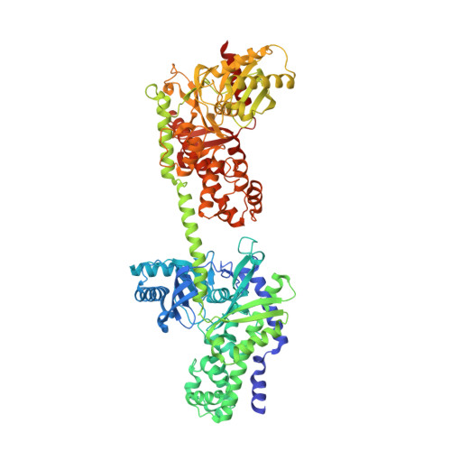
1DGK

MUTANT MONOMER OF RECOMBINANT HUMAN HEXOKINASE TYPE I WITH GLUCOSE AND ADP IN THE ACTIVE SITE


Experimental Data Snapshot

  • Method: X-RAY DIFFRACTION
  • Resolution: 2.80 Å
  • R-Value Free: 0.308 
  • R-Value Work: 0.258 
  • R-Value Observed: 0.260 

wwPDB Validation   3D Report Full Report


Ligand Structure Quality Assessment 


This is version 1.6 of the entry. See complete history


Literature

Crystal structures of mutant monomeric hexokinase I reveal multiple ADP binding sites and conformational changes relevant to allosteric regulation.

Aleshin, A.E.Kirby, C.Liu, X.Bourenkov, G.P.Bartunik, H.D.Fromm, H.J.Honzatko, R.B.

(2000) J Mol Biol 296: 1001-1015

  • DOI: https://doi.org/10.1006/jmbi.1999.3494
  • Primary Citation of Related Structures:  
    1CZA, 1DGK

  • PubMed Abstract: 

    Hexokinase I, the pacemaker of glycolysis in brain tissue, is composed of two structurally similar halves connected by an alpha-helix. The enzyme dimerizes at elevated protein concentrations in solution and in crystal structures; however, almost all published data reflect the properties of a hexokinase I monomer in solution. Crystal structures of mutant forms of recombinant human hexokinase I, presented here, reveal the enzyme monomer for the first time. The mutant hexokinases bind both glucose 6-phosphate and glucose with high affinity to their N and C-terminal halves, and ADP, also with high affinity, to a site near the N terminus of the polypeptide chain. Exposure of the monomer crystals to ADP in the complete absence of glucose 6-phosphate reveals a second binding site for adenine nucleotides at the putative active site (C-half), with conformational changes extending 15 A to the contact interface between the N and C-halves. The structures reveal distinct conformational states for the C-half and a rigid-body rotation of the N-half, as possible elements of a structure-based mechanism for allosteric regulation of catalysis.


  • Organizational Affiliation

    Department of Biochemistry Biophysics and Molecular Biology, Iowa State University, Ames, IA 50011, USA.


Macromolecules
Find similar proteins by:  (by identity cutoff)  |  3D Structure
Entity ID: 1
MoleculeChains Sequence LengthOrganismDetailsImage
HEXOKINASE TYPE IA [auth N]917Homo sapiensMutation(s): 4 
EC: 2.7.1.1
UniProt & NIH Common Fund Data Resources
Find proteins for P19367 (Homo sapiens)
Explore P19367 
Go to UniProtKB:  P19367
PHAROS:  P19367
GTEx:  ENSG00000156515 
Entity Groups  
Sequence Clusters30% Identity50% Identity70% Identity90% Identity95% Identity100% Identity
UniProt GroupP19367
Sequence Annotations
Expand
  • Reference Sequence
Experimental Data & Validation

Experimental Data

  • Method: X-RAY DIFFRACTION
  • Resolution: 2.80 Å
  • R-Value Free: 0.308 
  • R-Value Work: 0.258 
  • R-Value Observed: 0.260 
  • Space Group: P 21 21 2
Unit Cell:
Length ( Å )Angle ( ˚ )
a = 145.78α = 90
b = 146.83β = 90
c = 58.59γ = 90
Software Package:
Software NamePurpose
AMoREphasing
REFMACrefinement
DENZOdata reduction
SCALEPACKdata scaling

Structure Validation

View Full Validation Report


Ligand Structure Quality Assessment 


Entry History 

Deposition Data

Revision History  (Full details and data files)

  • Version 1.0: 2000-03-08
    Type: Initial release
  • Version 1.1: 2008-04-27
    Changes: Version format compliance
  • Version 1.2: 2011-07-13
    Changes: Version format compliance
  • Version 1.3: 2018-01-31
    Changes: Database references
  • Version 1.4: 2020-07-29
    Type: Remediation
    Reason: Carbohydrate remediation
    Changes: Data collection, Database references, Derived calculations, Structure summary
  • Version 1.5: 2021-11-03
    Changes: Database references, Structure summary
  • Version 1.6: 2024-02-07
    Changes: Data collection