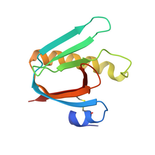
Exploration of the GM1 receptor-binding site of heat-labile enterotoxin and cholera toxin by phenyl-ring-containing galactose derivatives.
Fan, E., Merritt, E.A., Zhang, Z., Pickens, J.C., Roach, C., Ahn, M., Hol, W.G.(2001) Acta Crystallogr D Biol Crystallogr 57: 201-212
- PubMed: 11173465
- DOI: https://doi.org/10.1107/s0907444900016814
- Primary Citation of Related Structures:
1EEF, 1EEI, 1EFI, 1FD7 - PubMed Abstract:
Cholera toxin (CT) and the closely related heat-labile enterotoxin of Escherichia coli (LT) are responsible for numerous cases of diarrhea worldwide, leading to considerable morbidity and mortality. The B subunits of these heterohexameric AB(5) toxins form a pentameric arrangement which is responsible for binding to the receptor GM1 of the target epithelial cells of the host. Blocking these B pentamer-receptor interactions forms an avenue for therapeutic intervention. Here, the structural characterization of potential receptor-blocking compounds are described based on the previously identified inhibitor m-nitrophenyl-alpha-D-galactoside (MNPG). The structure of a CTB-MNPG complex confirms that the binding mode of this inhibitor is identical in the two homologous toxins CT and LT and is characterized by a glycosyl linkage geometry that leads to displacement of a well ordered water molecule near the amide group of Gly33 by the O1-substituent of MNPG. This glycosyl geometry is not maintained in the absence of a substituent that can displace this water, as shown by a complex of LTB with p-aminophenyl-alpha-D-galactoside (PAPG). New compounds were synthesized to investigate the feasibility of maintaining the favorable binding interactions exhibited by MNPG while gaining increased affinity through the addition of hydrophobic substituents complementary to either of two hydrophobic regions of the receptor-binding site. The structural characterization of complexes of LTB with two of these compounds, 3-benzylaminocarbonylphenyl-alpha-D-galactoside (BAPG) and 2-phenethyl-7-(2,3-dihydrophthalazine-1,4-dione)-alpha-D-galactoside (PEPG), demonstrates a partial success in this goal. Both compounds exhibit a mixture of binding modes, some of which are presumably influenced by the local packing environment at multiple crystallographically independent binding sites. The terminal phenyl ring of BAPG associates either with the phenyl group of Tyr12 or with the hydrophobic patch formed by Lys34 and Ile58. The latter interaction is also made by the terminal phenyl substituent of PEPG, despite a larger ring system linking the galactose moiety to the terminal phenyl. However, neither BAPG nor PEPG displaces the intended target water molecule. Both of the designed compounds exhibit increased affinity relative to the galactose and to PAPG notwithstanding the failure to displace a bound water, confirming that additional favorable hydrophobic interactions can be gained by extending the starting inhibitor by a hydrophobic tail. The insight gained from these structures should allow the design of additional candidate inhibitors that retain both the glycosyl geometry and water displacement exhibited by MNPG and the favorable hydrophobic interactions exhibited by BAPG and PEPG.
- Department of Biological Structure, University of Washington, Seattle 98195, USA.
Organizational Affiliation:


