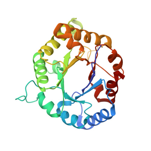
Crystal structure of recombinant human triosephosphate isomerase at 2.8 A resolution. Triosephosphate isomerase-related human genetic disorders and comparison with the trypanosomal enzyme.
Mande, S.C., Mainfroid, V., Kalk, K.H., Goraj, K., Martial, J.A., Hol, W.G.(1994) Protein Sci 3: 810-821
- PubMed: 8061610
- DOI: https://doi.org/10.1002/pro.5560030510
- Primary Citation of Related Structures:
1HTI - PubMed Abstract:
The crystal structure of recombinant human triosephosphate isomerase (hTIM) has been determined complexed with the transition-state analogue 2-phosphoglycolate at a resolution of 2.8 A. After refinement, the R-factor is 16.7% with good geometry. The asymmetric unit contains 1 complete dimer of 53,000 Da, with only 1 of the subunits binding the inhibitor. The so-called flexible loop, comprising residues 168-174, is in its "closed" conformation in the subunit that binds the inhibitor, and in the "open" conformation in the other subunit. The tips of the loop in these 2 conformations differ up to 7 A in position. The RMS difference between hTIM and the enzyme of Trypanosoma brucei, the causative agent of sleeping sickness, is 1.12 A for 487 C alpha positions with 53% sequence identity. Significant sequence differences between the human and parasite enzymes occur at about 13 A from the phosphate binding site. The chicken and human enzymes have an RMS difference of 0.69 A for 484 equivalent residues and about 90% sequence identity. Complementary mutations ensure a great similarity in the packing of side chains in the core of the beta-barrels of these 2 enzymes. Three point mutations in hTIM have been correlated with severe genetic disorders ranging from hemolytic disorder to neuromuscular impairment. Knowledge of the structure of the human enzyme provides insight into the probable effect of 2 of these mutations, Glu 104 to Asp and Phe 240 to Ile, on the enzyme. The third mutation reported to be responsible for a genetic disorder, Gly 122 to Arg, is however difficult to explain. This residue is far away from both catalytic centers in the dimer, as well as from the dimer interface, and seems unlikely to affect stability or activity. Inspection of the 3-dimensional structure of trypanosomal triosephosphate isomerase, which has a methionine at position 122, only increased the mystery of the effects of the Gly to Arg mutation in the human enzyme.
Organizational Affiliation:
Department of Biological Structure, School of Medicine, University of Washington, Seattle 98195.


