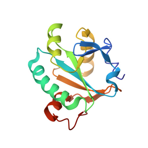
1I5G

TRYPAREDOXIN II COMPLEXED WITH GLUTATHIONYLSPERMIDINE


Experimental Data Snapshot

  • Method: X-RAY DIFFRACTION
  • Resolution: 1.40 Å
  • R-Value Free: 0.203 
  • R-Value Work: 0.168 
  • R-Value Observed: 0.170 

Starting Model: experimental
View more details

wwPDB Validation   3D Report Full Report


Ligand Structure Quality Assessment 


This is version 1.4 of the entry. See complete history


Literature

Structures of tryparedoxins revealing interaction with trypanothione.

Hofmann, B.Budde, H.Bruns, K.Guerrero, S.A.Kalisz, H.M.Menge, U.Montemartini, M.Nogoceke, E.Steinert, P.Wissing, J.B.Flohe, L.Hecht, H.J.

(2001) Biol Chem 382: 459-471

  • DOI: https://doi.org/10.1515/BC.2001.056
  • Primary Citation of Related Structures:  
    1EWX, 1EZK, 1FG4, 1I5G

  • PubMed Abstract: 

    Tryparedoxins (TXNs) catalyse the reduction of peroxiredoxin-type peroxidases by the bis-glutathionyl derivative of spermidine, trypanothione, and are relevant to hydroperoxide detoxification and virulence of trypanosomes. The 3D-structures of the following tryparedoxins are presented: authentic tryparedoxin1 of Crithidia fasciculata, CfTXN1; the his-tagged recombinant protein, CfTXN1H6; reduced and oxidised CfTXN2, and an alternative substrate derivative of the mutein CfTXN2H6-Cys44Ser. Cys41 (Cys40 in TXN1) of the active site motif 40-WCPPCR-45 proved to be the only solvent-exposed redox active residue in CfTXN2. In reduced TXNs, its nucleophilicity is increased by a network of hydrogen bonds. In oxidised TXNs it can be attacked by the thiol of the 1N-glutathionyl residue of trypanothione, as evidenced by the structure of 1N-glutathionylspermidine-derivatised CfTXN2H6-Cys44Ser. Modelling suggests Arg45 (44), Glu73 (72), the Ile110 (109) cis-Pro111 (110)-bond and Arg129 (128) to be involved in the binding of trypanothione to CfTXN2 (CfTXN1). The model of TXN-substrate interaction is consistent with functional characteristics of known and newly designed muteins (CfTXN2H6-Arg129Asp and Glu73Arg) and the 1N-glutathionyl-spermidine binding in the CfTXN2H6-Cys44Ser structure.


  • Organizational Affiliation

    Department of Biochemistry, Technical University of Braunschweig, Germany.


Macromolecules
Find similar proteins by:  (by identity cutoff)  |  3D Structure
Entity ID: 1
MoleculeChains Sequence LengthOrganismDetailsImage
TRYPAREDOXIN II144Crithidia fasciculataMutation(s): 1 
EC: 1.8.1.8
UniProt
Find proteins for O77093 (Crithidia fasciculata)
Explore O77093 
Go to UniProtKB:  O77093
Entity Groups  
Sequence Clusters30% Identity50% Identity70% Identity90% Identity95% Identity100% Identity
UniProt GroupO77093
Sequence Annotations
Expand
  • Reference Sequence
Small Molecules
Ligands 2 Unique
IDChains Name / Formula / InChI Key2D Diagram3D Interactions
TS5
Query on TS5

Download Ideal Coordinates CCD File 
C [auth A]GLUTATHIONYLSPERMIDINE
C17 H34 N6 O5 S
NEDQLXHBVHSKNV-STQMWFEESA-N
TRS
Query on TRS

Download Ideal Coordinates CCD File 
B [auth A]2-AMINO-2-HYDROXYMETHYL-PROPANE-1,3-DIOL
C4 H12 N O3
LENZDBCJOHFCAS-UHFFFAOYSA-O
Experimental Data & Validation

Experimental Data

  • Method: X-RAY DIFFRACTION
  • Resolution: 1.40 Å
  • R-Value Free: 0.203 
  • R-Value Work: 0.168 
  • R-Value Observed: 0.170 
  • Space Group: P 21 21 21
Unit Cell:
Length ( Å )Angle ( ˚ )
a = 41.211α = 90
b = 50.51β = 90
c = 67.259γ = 90
Software Package:
Software NamePurpose
AMoREphasing
REFMACrefinement
MOSFLMdata reduction
CCP4data scaling

Structure Validation

View Full Validation Report


Ligand Structure Quality Assessment 


Entry History 

Revision History  (Full details and data files)

  • Version 1.0: 2001-03-14
    Type: Initial release
  • Version 1.1: 2008-04-27
    Changes: Version format compliance
  • Version 1.2: 2011-07-13
    Changes: Version format compliance
  • Version 1.3: 2021-10-27
    Changes: Database references, Derived calculations
  • Version 1.4: 2023-08-09
    Changes: Data collection, Refinement description