1PQY

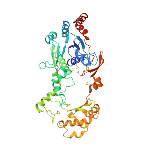
Crystal structure of formyl-coA transferase yfdW from E. coli


Experimental Data Snapshot

  • Method: X-RAY DIFFRACTION
  • Resolution: 2.35 Å
  • R-Value Free: 0.211 
  • R-Value Work: 0.177 

wwPDB Validation   3D Report Full Report


This is version 1.4 of the entry. See complete history


Literature

Structure of Escherichia coli YfdW, a type III CoA transferase.

Gogos, A.Gorman, J.Shapiro, L.

(2004) Acta Crystallogr D Biol Crystallogr 60: 507-511

  • DOI: https://doi.org/10.1107/S0907444904000034
  • Primary Citation of Related Structures:  
    1PQY, 1Q6Y, 1Q7E

  • PubMed Abstract: 

    Crystal structures are reported for free and coenzyme A (CoA) bound forms of the YfdW protein from Escherichia coli, a representative type III CoA transferase. The structures reveal a two-domain protomer with interdomain connections forming a ring-like structure with a large central hole. Two protomers associate to form a highly intertwined dimer in which the hole of each ring is filled by the partner molecule. Each protomer binds a single CoA molecule and these CoA-binding sites are distant from one another in the dimer.


  • Organizational Affiliation

    Department of Biochemistry and Molecular Biophysics, Columbia University College of Physicians and Surgeons, 630 West 168th Street, New York, NY 10032, USA.


Macromolecules
Find similar proteins by:  (by identity cutoff)  |  3D Structure
Entity ID: 1
MoleculeChains Sequence LengthOrganismDetailsImage
Hypothetical protein yfdW428Escherichia coliMutation(s): 10 
Gene Names: YFDW
EC: 2.8.3.16
UniProt
Find proteins for P69902 (Escherichia coli (strain K12))
Explore P69902 
Go to UniProtKB:  P69902
Entity Groups  
Sequence Clusters30% Identity50% Identity70% Identity90% Identity95% Identity100% Identity
UniProt GroupP69902
Sequence Annotations
Expand
  • Reference Sequence
Small Molecules
Modified Residues  1 Unique
IDChains TypeFormula2D DiagramParent
MSE
Query on MSE
A
L-PEPTIDE LINKINGC5 H11 N O2 SeMET
Experimental Data & Validation

Experimental Data

  • Method: X-RAY DIFFRACTION
  • Resolution: 2.35 Å
  • R-Value Free: 0.211 
  • R-Value Work: 0.177 
  • Space Group: C 1 2 1
Unit Cell:
Length ( Å )Angle ( ˚ )
a = 92.261α = 90
b = 69.539β = 108.64
c = 73.109γ = 90
Software Package:
Software NamePurpose
CNSrefinement
DENZOdata reduction
SCALEPACKdata scaling
SHARPphasing

Structure Validation

View Full Validation Report


Entry History 

Deposition Data

Revision History  (Full details and data files)

  • Version 1.0: 2003-09-30
    Type: Initial release
  • Version 1.1: 2008-04-29
    Changes: Version format compliance
  • Version 1.2: 2011-07-13
    Changes: Derived calculations, Version format compliance
  • Version 1.3: 2021-02-03
    Changes: Database references, Derived calculations, Structure summary
  • Version 1.4: 2024-10-30
    Changes: Data collection, Database references, Structure summary