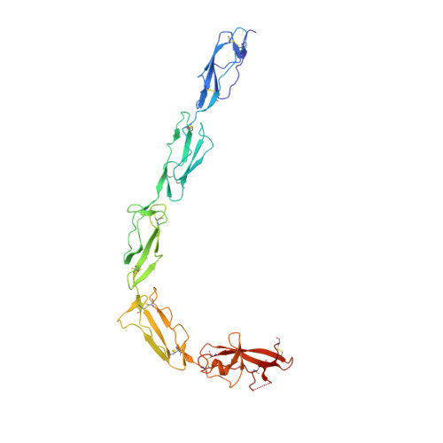
1QUB

CRYSTAL STRUCTURE OF THE GLYCOSYLATED FIVE-DOMAIN HUMAN BETA2-GLYCOPROTEIN I PURIFIED FROM BLOOD PLASMA


Experimental Data Snapshot

  • Method: X-RAY DIFFRACTION
  • Resolution: 2.70 Å
  • R-Value Free: 0.269 
  • R-Value Work: 0.249 
  • R-Value Observed: 0.250 

wwPDB Validation   3D Report Full Report


Ligand Structure Quality Assessment 


This is version 2.1 of the entry. See complete history


Literature

Adhesion mechanism of human beta(2)-glycoprotein I to phospholipids based on its crystal structure.

Bouma, B.de Groot, P.G.van den Elsen, J.M.Ravelli, R.B.Schouten, A.Simmelink, M.J.Derksen, R.H.Kroon, J.Gros, P.

(1999) EMBO J 18: 5166-5174

  • DOI: https://doi.org/10.1093/emboj/18.19.5166
  • Primary Citation of Related Structures:  
    1QUB

  • PubMed Abstract: 

    Human beta(2)-glycoprotein I is a heavily glycosylated five-domain plasma membrane-adhesion protein, which has been implicated in blood coagulation and clearance of apoptotic bodies from the circulation. It is also the key antigen in the autoimmune disease anti-phospholipid syndrome. The crystal structure of beta(2)-glycoprotein I isolated from human plasma reveals an elongated fish-hook-like arrangement of the globular short consensus repeat domains. Half of the C-terminal fifth domain deviates strongly from the standard fold, as observed in domains one to four. This aberrant half forms a specific phospholipid-binding site. A large patch of 14 positively charged residues provides electrostatic interactions with anionic phospholipid headgroups and an exposed membrane-insertion loop yields specificity for lipid layers. The observed spatial arrangement of the five domains suggests a functional partitioning of protein adhesion and membrane adhesion over the N- and C-terminal domains, respectively, separated by glycosylated bridging domains. Coordinates are in the Protein Data Bank (accession No. 1QUB).


  • Organizational Affiliation

    Department of Crystal and Structural Chemistry, Bijvoet Center for Biomolecular Research, Utrecht University, Padualaan 8, 3584 CH Utrecht, The Netherlands.


Macromolecules
Find similar proteins by:  (by identity cutoff)  |  3D Structure
Entity ID: 1
MoleculeChains Sequence LengthOrganismDetailsImage
PROTEIN (human beta2-Glycoprotein I)319Homo sapiensMutation(s): 0 
UniProt & NIH Common Fund Data Resources
Find proteins for P02749 (Homo sapiens)
Explore P02749 
Go to UniProtKB:  P02749
PHAROS:  P02749
GTEx:  ENSG00000091583 
Entity Groups  
Sequence Clusters30% Identity50% Identity70% Identity90% Identity95% Identity100% Identity
UniProt GroupP02749
Glycosylation
Glycosylation Sites: 4Go to GlyGen: P02749-1
Sequence Annotations
Expand
  • Reference Sequence
Oligosaccharides

Help

Entity ID: 2
MoleculeChains Length2D Diagram Glycosylation3D Interactions
alpha-D-mannopyranose-(1-4)-2-acetamido-2-deoxy-beta-D-glucopyranose-(1-4)-2-acetamido-2-deoxy-beta-D-glucopyranose
B
3N-Glycosylation
Glycosylation Resources
GlyTouCan:  G62182OO
GlyCosmos:  G62182OO
GlyGen:  G62182OO
Entity ID: 3
MoleculeChains Length2D Diagram Glycosylation3D Interactions
2-acetamido-2-deoxy-beta-D-glucopyranose-(1-4)-2-acetamido-2-deoxy-beta-D-glucopyranose
C
2N-Glycosylation
Glycosylation Resources
GlyTouCan:  G42666HT
GlyCosmos:  G42666HT
GlyGen:  G42666HT
Small Molecules
Ligands 1 Unique
IDChains Name / Formula / InChI Key2D Diagram3D Interactions
NAG
Query on NAG

Download Ideal Coordinates CCD File 
D [auth A],
E [auth A]
2-acetamido-2-deoxy-beta-D-glucopyranose
C8 H15 N O6
OVRNDRQMDRJTHS-FMDGEEDCSA-N
Experimental Data & Validation

Experimental Data

  • Method: X-RAY DIFFRACTION
  • Resolution: 2.70 Å
  • R-Value Free: 0.269 
  • R-Value Work: 0.249 
  • R-Value Observed: 0.250 
  • Space Group: C 2 2 21
Unit Cell:
Length ( Å )Angle ( ˚ )
a = 161.17α = 90
b = 166.49β = 90
c = 114.51γ = 90
Software Package:
Software NamePurpose
MOSFLMdata reduction
TRUNCATEdata reduction
SOLVEphasing
CNSrefinement
CCP4data scaling

Structure Validation

View Full Validation Report


Ligand Structure Quality Assessment 


Entry History 

Deposition Data

Revision History  (Full details and data files)

  • Version 1.0: 1999-10-08
    Type: Initial release
  • Version 1.1: 2008-04-27
    Changes: Version format compliance
  • Version 1.2: 2011-07-13
    Changes: Non-polymer description, Version format compliance
  • Version 1.3: 2018-01-24
    Changes: Structure summary
  • Version 2.0: 2020-07-29
    Type: Remediation
    Reason: Carbohydrate remediation
    Changes: Advisory, Atomic model, Data collection, Derived calculations, Structure summary
  • Version 2.1: 2024-10-30
    Changes: Data collection, Database references, Structure summary