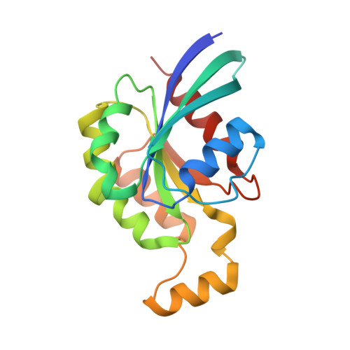
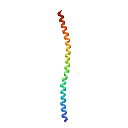
1S1C

Crystal structure of the complex between the human RhoA and Rho-binding domain of human ROCKI


Experimental Data Snapshot

  • Method: X-RAY DIFFRACTION
  • Resolution: 2.60 Å
  • R-Value Free: 0.257 
  • R-Value Work: 0.219 
  • R-Value Observed: 0.219 

wwPDB Validation   3D Report Full Report


Ligand Structure Quality Assessment 


This is version 1.3 of the entry. See complete history


Literature

Structural Insights into the Interaction of ROCKI with the Switch Regions of RhoA.

Dvorsky, R.Blumenstein, L.Vetter, I.R.Ahmadian, M.R.

(2004) J Biol Chem 279: 7098-7104

  • DOI: https://doi.org/10.1074/jbc.M311911200
  • Primary Citation of Related Structures:  
    1S1C

  • PubMed Abstract: 

    The Rho-ROCK pathway modulates the phosphorylation level of a variety of important signaling proteins and is thereby involved in miscellaneous cellular processes including cell migration, neurite outgrowth, and smooth muscle contraction. The observation of the involvement of the Rho-ROCK pathway in tumor invasion and in diseases such as hypertension and bronchial asthma makes it an interesting target for drug development. We herein present the crystal structure of the complex between active RhoA and the Rho-binding domain of ROCKI. The Rho-binding domain structure forms a parallel alpha-helical coiled-coil dimer and, in contrast to the published Rho-protein kinase N structure, binds exclusively to the switch I and II regions of the guanosine 5'-(beta,gamma-imido)triphosphate-bound RhoA. The switch regions of two different RhoA molecules form a predominantly hydrophobic patch, which is complementarily bound by two identical short helices of 13 residues (amino acids 998-1010). The identified ROCK-binding site of RhoA strikingly supports the assumption of a common consensus-binding site for effector recognition.


  • Organizational Affiliation

    Max-Planck-Institute fuer Molekulare Physiologie, Abteilung Strukturelle Biologie, Otto-Hahn-Strasse 11, 44227 Dortmund, Germany.


Macromolecules
Find similar proteins by:  (by identity cutoff)  |  3D Structure
Entity ID: 1
MoleculeChains Sequence LengthOrganismDetailsImage
Transforming protein RhoA
A, B
183Homo sapiensMutation(s): 0 
Gene Names: ARHAARH12RHOARHO12
EC: 3.6.5.2
UniProt & NIH Common Fund Data Resources
Find proteins for P61586 (Homo sapiens)
Explore P61586 
Go to UniProtKB:  P61586
PHAROS:  P61586
GTEx:  ENSG00000067560 
Entity Groups  
Sequence Clusters30% Identity50% Identity70% Identity90% Identity95% Identity100% Identity
UniProt GroupP61586
Sequence Annotations
Expand
  • Reference Sequence
Find similar proteins by:  (by identity cutoff)  |  3D Structure
Entity ID: 2
MoleculeChains Sequence LengthOrganismDetailsImage
Rho-associated, coiled-coil containing protein kinase 1C [auth X],
D [auth Y]
71Homo sapiensMutation(s): 0 
EC: 2.7.11.1
UniProt & NIH Common Fund Data Resources
Find proteins for Q13464 (Homo sapiens)
Explore Q13464 
Go to UniProtKB:  Q13464
PHAROS:  Q13464
GTEx:  ENSG00000067900 
Entity Groups  
Sequence Clusters30% Identity50% Identity70% Identity90% Identity95% Identity100% Identity
UniProt GroupQ13464
Sequence Annotations
Expand
  • Reference Sequence
Experimental Data & Validation

Experimental Data

  • Method: X-RAY DIFFRACTION
  • Resolution: 2.60 Å
  • R-Value Free: 0.257 
  • R-Value Work: 0.219 
  • R-Value Observed: 0.219 
  • Space Group: P 1 21 1
Unit Cell:
Length ( Å )Angle ( ˚ )
a = 34.404α = 90
b = 89.537β = 91.86
c = 98.105γ = 90
Software Package:
Software NamePurpose
CNSrefinement
DENZOdata reduction
SCALEPACKdata scaling
AMoREphasing

Structure Validation

View Full Validation Report


Ligand Structure Quality Assessment 


Entry History 

Deposition Data

Revision History  (Full details and data files)

  • Version 1.0: 2004-02-10
    Type: Initial release
  • Version 1.1: 2008-04-29
    Changes: Version format compliance
  • Version 1.2: 2011-07-13
    Changes: Version format compliance
  • Version 1.3: 2024-02-14
    Changes: Data collection, Database references, Derived calculations