1U9Z

Crystal Structure of Phosphoribosyl Diphosphate Synthase Complexed with AMP and Ribose 5-Phosphate


Experimental Data Snapshot

  • Method: X-RAY DIFFRACTION
  • Resolution: 2.80 Å
  • R-Value Free: 0.272 
  • R-Value Work: 0.238 
  • R-Value Observed: 0.240 

Starting Model: experimental
View more details

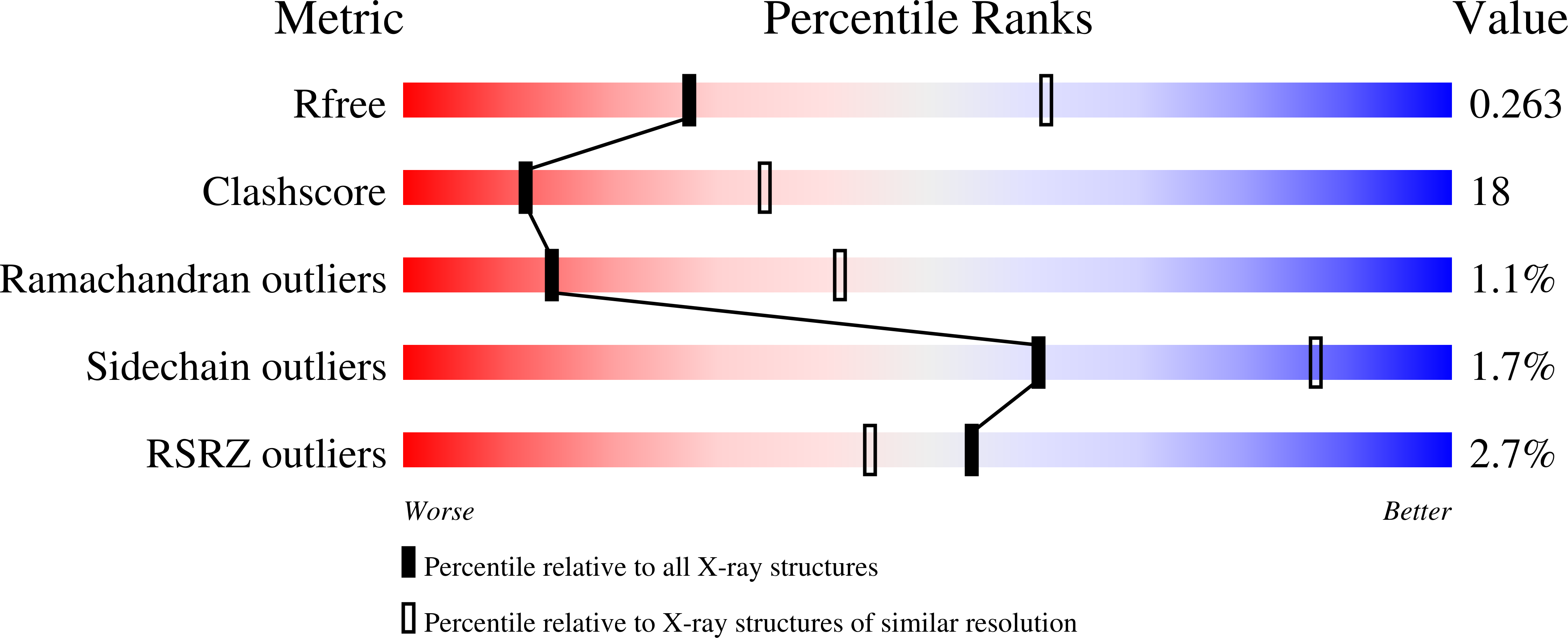
wwPDB Validation   3D Report Full Report


Ligand Structure Quality Assessment 


This is version 1.4 of the entry. See complete history


Literature

Novel class III phosphoribosyl diphosphate synthase: structure and properties of the tetrameric, phosphate-activated, non-allosterically inhibited enzyme from Methanocaldococcus jannaschii

Kadziola, A.Jepsen, C.H.Johansson, E.McGuire, J.Larsen, S.Hove-Jensen, B.

(2005) J Mol Biol 354: 815-828

  • DOI: https://doi.org/10.1016/j.jmb.2005.10.001
  • Primary Citation of Related Structures:  
    1U9Y, 1U9Z

  • PubMed Abstract: 

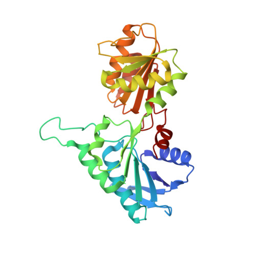
    The prs gene encoding phosphoribosyl diphosphate (PRPP) synthase of the hyperthermophilic autotrophic methanogenic archaeon Methanocaldococcus jannaschii has been cloned and expressed in Escherichia coli. Subsequently, M.jannaschii PRPP synthase has been purified, characterised, crystallised, and the crystal structure determined. The enzyme is activated by phosphate ions and only ATP or dATP serve as diphosphoryl donors. The K(m) values are determined as 2.6 mM and 2.8 mM for ATP and ribose 5-phosphate, respectively, and the V(max) value as 2.20 mmol (minxmg of protein)(-1). ADP is a potent inhibitor of activity while GDP has no effect. A single ADP binding site, the active site, is present per subunit. The crystal structure of the enzyme reveals a more compact subunit than that of the enzyme from the mesophile Bacillus subtilis, caused by truncations at the N and C terminus as well as shorter loops in the M.jannaschii enzyme. The M.jannaschii enzyme displays a tetrameric quaternary structure in contrast to the hexameric quaternary structure of B.subtilis PRPP synthase. Soaking of the crystals with 5'-AMP and PRPP revealed the position of the former compound as well as that of ribose 5-phosphate. The properties of M.jannaschii PRPP synthase differ widely from previously characterised PRPP synthases by its tetrameric quaternary structure and the simultaneous phosphate ion-activation and lack of allosteric inhibition, and, thus, constitute a novel class of PRPP synthases.


  • Organizational Affiliation

    Centre for Crystallographic Studies, Department of Chemistry, University of Copenhagen, 5 Universitetsparken, DK-2100 Copenhagen Ø, Denmark. anders@ccs.ki.ku.dk


Macromolecules
Find similar proteins by:  (by identity cutoff)  |  3D Structure
Entity ID: 1
MoleculeChains Sequence LengthOrganismDetailsImage
Ribose-phosphate pyrophosphokinase
A, B, C, D
284Methanocaldococcus jannaschiiMutation(s): 0 
Gene Names: prs
EC: 2.7.6.1
UniProt
Find proteins for Q58761 (Methanocaldococcus jannaschii (strain ATCC 43067 / DSM 2661 / JAL-1 / JCM 10045 / NBRC 100440))
Explore Q58761 
Go to UniProtKB:  Q58761
Entity Groups  
Sequence Clusters30% Identity50% Identity70% Identity90% Identity95% Identity100% Identity
UniProt GroupQ58761
Sequence Annotations
Expand
  • Reference Sequence
Small Molecules
Ligands 2 Unique
IDChains Name / Formula / InChI Key2D Diagram3D Interactions
AMP
Query on AMP

Download Ideal Coordinates CCD File 
G [auth B],
H [auth B],
J [auth C],
L [auth D]
ADENOSINE MONOPHOSPHATE
C10 H14 N5 O7 P
UDMBCSSLTHHNCD-KQYNXXCUSA-N
R5P
Query on R5P

Download Ideal Coordinates CCD File 
E [auth A],
F [auth B],
I [auth C],
K [auth D]
RIBOSE-5-PHOSPHATE
C5 H11 O8 P
PPQRONHOSHZGFQ-LMVFSUKVSA-N
Experimental Data & Validation

Experimental Data

  • Method: X-RAY DIFFRACTION
  • Resolution: 2.80 Å
  • R-Value Free: 0.272 
  • R-Value Work: 0.238 
  • R-Value Observed: 0.240 
  • Space Group: P 21 21 21
Unit Cell:
Length ( Å )Angle ( ˚ )
a = 71.26α = 90
b = 138.25β = 90
c = 138.4γ = 90
Software Package:
Software NamePurpose
DENZOdata reduction
SCALEPACKdata scaling
AMoREphasing
CNSrefinement

Structure Validation

View Full Validation Report


Ligand Structure Quality Assessment 


Entry History 

Deposition Data

Revision History  (Full details and data files)

  • Version 1.0: 2005-08-23
    Type: Initial release
  • Version 1.1: 2008-04-30
    Changes: Version format compliance
  • Version 1.2: 2011-07-13
    Changes: Version format compliance
  • Version 1.3: 2018-03-07
    Changes: Data collection
  • Version 1.4: 2023-08-23
    Changes: Data collection, Database references, Derived calculations, Refinement description