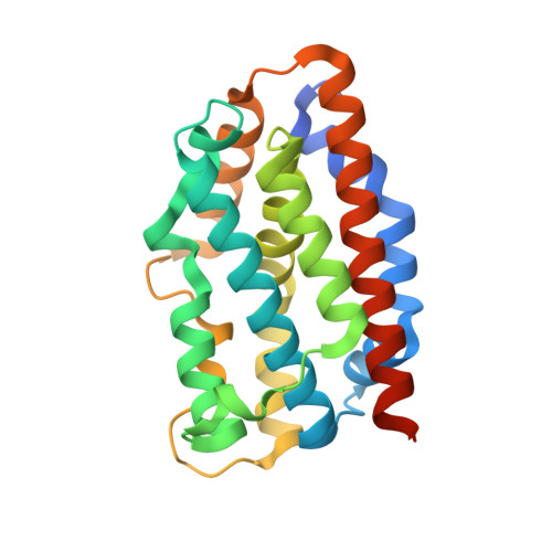
1XK0

Crystal Structures of the G139A, G139A-NO and G143H Mutants of Human Heme Oxygenase-1


Experimental Data Snapshot

  • Method: X-RAY DIFFRACTION
  • Resolution: 2.18 Å
  • R-Value Free: 0.266 
  • R-Value Work: 0.224 

wwPDB Validation   3D Report Full Report


Ligand Structure Quality Assessment 


This is version 1.4 of the entry. See complete history


Literature

Crystal structures of the G139A, G139A-NO and G143H mutants of human heme oxygenase-1. A finely tuned hydrogen-bonding network controls oxygenase versus peroxidase activity.

Lad, L.Koshkin, A.Ortiz de Montellano, P.R.Poulos, T.L.

(2005) J Biol Inorg Chem 10: 138-146

  • DOI: https://doi.org/10.1007/s00775-004-0620-6
  • Primary Citation of Related Structures:  
    1XJZ, 1XK0, 1XK1

  • PubMed Abstract: 

    Conserved glycines, Gly139 and Gly143, in the distal helix of human heme oxygenase-1 (HO-1) provide the flexibility required for the opening and closing of the heme active site for substrate binding and product dissociation during HO-1 catalysis. Earlier mutagenesis work on human HO-1 showed that replacement of either Gly139 or Gly143 suppresses heme oxygenase activity and, in the case of the Gly139 mutants, increases peroxidase activity (Liu et al. in J. Biol. Chem. 275:34501, 2000). To further investigate the role of the conserved distal helix glycines, we have determined the crystal structures of the human HO-1 G139A mutant, the G139A mutant in a complex with NO, and the G143H mutant at 1.88, 2.18 and 2.08 A, respectively. The results confirm that fine tuning of the previously noted active-site hydrogen-bonding network is critical in determining whether heme oxygenase or peroxidase activity is observed.


  • Organizational Affiliation

    Department of Molecular Biology and Biochemistry, University of California, Irvine, 92697-3900, USA.


Macromolecules
Find similar proteins by:  (by identity cutoff)  |  3D Structure
Entity ID: 1
MoleculeChains Sequence LengthOrganismDetailsImage
Heme oxygenase 1
A, B
233Homo sapiensMutation(s): 1 
Gene Names: HMOX1HO1HO
EC: 1.14.99.3 (PDB Primary Data), 1.14.14.18 (UniProt)
UniProt & NIH Common Fund Data Resources
Find proteins for P09601 (Homo sapiens)
Explore P09601 
Go to UniProtKB:  P09601
PHAROS:  P09601
GTEx:  ENSG00000100292 
Entity Groups  
Sequence Clusters30% Identity50% Identity70% Identity90% Identity95% Identity100% Identity
UniProt GroupP09601
Sequence Annotations
Expand
  • Reference Sequence
Experimental Data & Validation

Experimental Data

  • Method: X-RAY DIFFRACTION
  • Resolution: 2.18 Å
  • R-Value Free: 0.266 
  • R-Value Work: 0.224 
  • Space Group: P 21 21 21
Unit Cell:
Length ( Å )Angle ( ˚ )
a = 54.06α = 90
b = 75.968β = 90
c = 105.299γ = 90
Software Package:
Software NamePurpose
HKL-2000data collection
SCALEPACKdata scaling
CNSrefinement
HKL-2000data reduction
CNSphasing

Structure Validation

View Full Validation Report


Ligand Structure Quality Assessment 


Entry History 

Deposition Data

Revision History  (Full details and data files)

  • Version 1.0: 2005-12-06
    Type: Initial release
  • Version 1.1: 2008-04-30
    Changes: Version format compliance
  • Version 1.2: 2011-07-13
    Changes: Version format compliance
  • Version 1.3: 2021-10-20
    Changes: Database references, Derived calculations
  • Version 1.4: 2024-02-14
    Changes: Data collection