1XO2

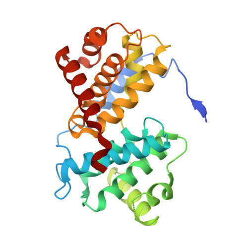
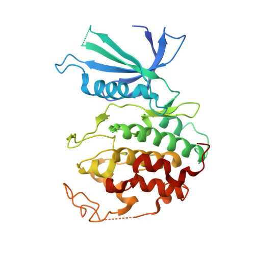
Crystal structure of a human cyclin-dependent kinase 6 complex with a flavonol inhibitor, fisetin


Experimental Data Snapshot

  • Method: X-RAY DIFFRACTION
  • Resolution: 2.90 Å
  • R-Value Free: 0.313 
  • R-Value Work: 0.260 

Starting Model: experimental
View more details

wwPDB Validation   3D Report Full Report


Ligand Structure Quality Assessment 


This is version 1.3 of the entry. See complete history


Literature

Crystal Structure of a Human Cyclin-Dependent Kinase 6 Complex with a Flavonol Inhibitor, Fisetin.

Lu, H.S.Chang, D.J.Baratte, B.Meijer, L.Schulze-Gahmen, U.

(2005) J Med Chem 48: 737-747

  • DOI: https://doi.org/10.1021/jm049353p
  • Primary Citation of Related Structures:  
    1XO2

  • PubMed Abstract: 

    Cyclin-dependent kinases (CDKs) play a central role in cell cycle control, apoptosis, transcription, and neuronal functions. They are important targets for the design of drugs with antimitotic or antineurodegenerative effects. CDK4 and CDK6 form a subfamily among the CDKs in mammalian cells, as defined by sequence similarities. Compared to CDK2 and CDK5, structural information on CDK4 and CDK6 is sparse. We describe here the crystal structure of human CDK6 in complex with a viral cyclin and a flavonol inhibitor, fisetin. Fisetin binds to the active form of CDK6, forming hydrogen bonds with the side chains of residues in the binding pocket that undergo large conformational changes during CDK activation by cyclin binding. The 4-keto group and the 3-hydroxyl group of fisetin are hydrogen bonded with the backbone in the hinge region between the N-terminal and C-terminal kinase domain, as has been observed for many CDK inhibitors. However, CDK2 and HCK kinase in complex with other flavone inhibitors such as quercetin and flavopiridol showed a different binding mode with the inhibitor rotated by about 180 degrees. The structural information of the CDK6-fisetin complex is correlated with the binding affinities of different flavone inhibitors for CDK6. This complex structure is the first description of an inhibitor complex with a kinase from the CDK4/6 subfamily and can provide a basis for selecting and designing inhibitor compounds with higher affinities and specificities.


  • Organizational Affiliation

    Physical Biosciences Division, Lawrence Berkeley National Laboratory, 1 Cyclotron Road, MS 64R0121, Berkeley, California 94720, USA.


Macromolecules
Find similar proteins by:  (by identity cutoff)  |  3D Structure
Entity ID: 1
MoleculeChains Sequence LengthOrganismDetailsImage
Cyclin254Saimiriine gammaherpesvirus 2Mutation(s): 0 
Gene Names: 72ECLF2
UniProt
Find proteins for Q01043 (Saimiriine herpesvirus 2 (strain 11))
Explore Q01043 
Go to UniProtKB:  Q01043
Entity Groups  
Sequence Clusters30% Identity50% Identity70% Identity90% Identity95% Identity100% Identity
UniProt GroupQ01043
Sequence Annotations
Expand
  • Reference Sequence
Find similar proteins by:  (by identity cutoff)  |  3D Structure
Entity ID: 2
MoleculeChains Sequence LengthOrganismDetailsImage
Cell division protein kinase 6308Homo sapiensMutation(s): 0 
Gene Names: CDK6
EC: 2.7.1.37 (PDB Primary Data), 2.7.11.22 (UniProt)
UniProt & NIH Common Fund Data Resources
Find proteins for Q00534 (Homo sapiens)
Explore Q00534 
Go to UniProtKB:  Q00534
PHAROS:  Q00534
GTEx:  ENSG00000105810 
Entity Groups  
Sequence Clusters30% Identity50% Identity70% Identity90% Identity95% Identity100% Identity
UniProt GroupQ00534
Sequence Annotations
Expand
  • Reference Sequence
Small Molecules
Ligands 1 Unique
IDChains Name / Formula / InChI Key2D Diagram3D Interactions
FSE
Query on FSE

Download Ideal Coordinates CCD File 
C [auth B]3,7,3',4'-TETRAHYDROXYFLAVONE
C15 H10 O6
XHEFDIBZLJXQHF-UHFFFAOYSA-N
Binding Affinity Annotations 
IDSourceBinding Affinity
FSE PDBBind:  1XO2 IC50: 850 (nM) from 1 assay(s)
BindingDB:  1XO2 IC50: 850 (nM) from 1 assay(s)
Experimental Data & Validation

Experimental Data

  • Method: X-RAY DIFFRACTION
  • Resolution: 2.90 Å
  • R-Value Free: 0.313 
  • R-Value Work: 0.260 
  • Space Group: P 65 2 2
Unit Cell:
Length ( Å )Angle ( ˚ )
a = 66.162α = 90
b = 66.162β = 90
c = 448.596γ = 120
Software Package:
Software NamePurpose
HKL-2000data collection
TRUNCATEdata reduction
AMoREphasing
CNSrefinement
HKL-2000data reduction
CCP4data scaling

Structure Validation

View Full Validation Report


Ligand Structure Quality Assessment 


Entry History 

Deposition Data

Revision History  (Full details and data files)

  • Version 1.0: 2005-03-01
    Type: Initial release
  • Version 1.1: 2008-04-30
    Changes: Version format compliance
  • Version 1.2: 2011-07-13
    Changes: Non-polymer description, Version format compliance
  • Version 1.3: 2023-08-23
    Changes: Data collection, Database references, Derived calculations, Refinement description, Structure summary