2B2N

Structure of transcription-repair coupling factor


Experimental Data Snapshot

  • Method: X-RAY DIFFRACTION
  • Resolution: 2.10 Å
  • R-Value Free: 0.232 
  • R-Value Work: 0.198 

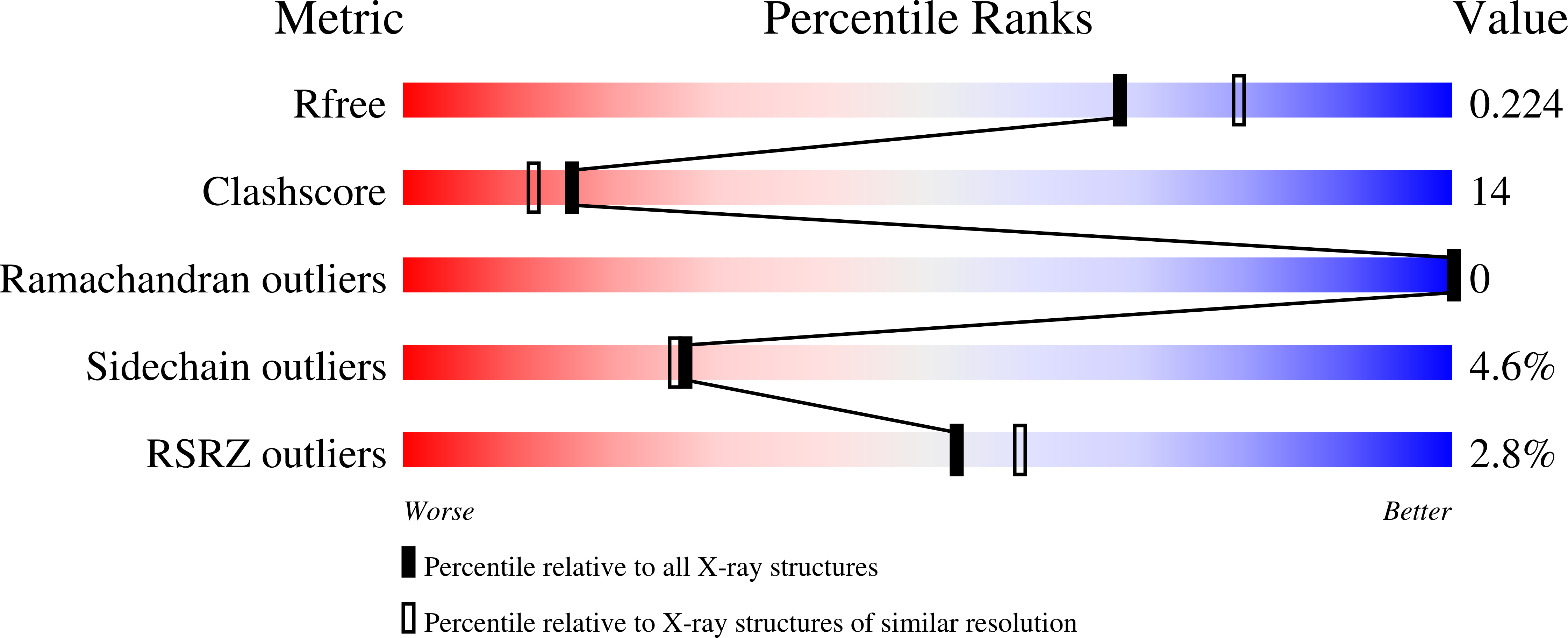
wwPDB Validation   3D Report Full Report


Ligand Structure Quality Assessment 


This is version 1.4 of the entry. See complete history


Literature

Structural Basis for Transcription-coupled Repair: the N Terminus of Mfd Resembles UvrB with Degenerate ATPase Motifs

Assenmacher, N.Wenig, K.Lammens, A.Hopfner, K.-P.

(2006) J Mol Biol 355: 675-683

  • DOI: https://doi.org/10.1016/j.jmb.2005.10.033
  • Primary Citation of Related Structures:  
    2B2N

  • PubMed Abstract: 

    The transcription repair coupling factor Mfd removes stalled RNA polymerase from DNA lesions and links transcription to UvrABC-dependent nucleotide excision repair in prokaryotes. We report the 2.1A crystal structure of the UvrA-binding N terminus (residues 1-333) of Escherichia coli Mfd (Mfd-N). Remarkably, Mfd-N reveals a fold that resembles the three N-terminal domains of the repair enzyme UvrB. Domain 1A of Mfd adopts a typical RecA fold, domain 1B matches the damage-binding domain of the UvrB, and domain 2 highly resembles the implicated UvrA-binding domain of UvrB. However, Mfd apparently lacks a functional ATP-binding site and does not contain the DNA damage-binding motifs of UvrB. Thus, our results suggest that Mfd might form a UvrA recruitment factor at stalled transcription complexes that architecturally but not catalytically resembles UvrB.


  • Organizational Affiliation

    Gene Center and Department of Chemistry and Biochemistry, University of Munich (LMU), Feodor-Lynen-Str. 25, D-81377 Munich, Germany.


Macromolecules
Find similar proteins by:  (by identity cutoff)  |  3D Structure
Entity ID: 1
MoleculeChains Sequence LengthOrganismDetailsImage
Transcription-repair coupling factor
A, B
344Escherichia coli K-12Mutation(s): 0 
Gene Names: mfd
EC: 3.6.4
UniProt
Find proteins for P30958 (Escherichia coli (strain K12))
Explore P30958 
Go to UniProtKB:  P30958
Entity Groups  
Sequence Clusters30% Identity50% Identity70% Identity90% Identity95% Identity100% Identity
UniProt GroupP30958
Sequence Annotations
Expand
  • Reference Sequence
Experimental Data & Validation

Experimental Data

  • Method: X-RAY DIFFRACTION
  • Resolution: 2.10 Å
  • R-Value Free: 0.232 
  • R-Value Work: 0.198 
  • Space Group: P 65 2 2
Unit Cell:
Length ( Å )Angle ( ˚ )
a = 112.65α = 90
b = 112.65β = 90
c = 213.86γ = 120
Software Package:
Software NamePurpose
ProDCdata collection
XDSdata reduction
SOLVEphasing
CNSrefinement
XDSdata scaling

Structure Validation

View Full Validation Report


Ligand Structure Quality Assessment 


Entry History 

Deposition Data

Revision History  (Full details and data files)

  • Version 1.0: 2006-01-24
    Type: Initial release
  • Version 1.1: 2008-05-01
    Changes: Version format compliance
  • Version 1.2: 2011-07-13
    Changes: Source and taxonomy, Version format compliance
  • Version 1.3: 2017-10-11
    Changes: Advisory, Refinement description
  • Version 1.4: 2024-02-14
    Changes: Advisory, Data collection, Database references, Derived calculations