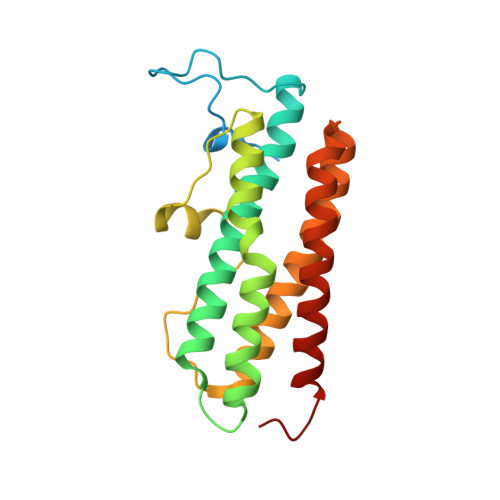
2C2F

Dps from Deinococcus radiodurans


Experimental Data Snapshot

  • Method: X-RAY DIFFRACTION
  • Resolution: 1.61 Å
  • R-Value Free: 0.153 
  • R-Value Work: 0.131 
  • R-Value Observed: 0.132 

wwPDB Validation   3D Report Full Report


This is version 1.2 of the entry. See complete history


Literature

The Crystal Structure of Deinococcus Radiodurans Dps Protein (Dr2263) Reveals the Presence of a Novel Metal Centre in the N Terminus.

Romao, C.V.Mitchell, E.Mcsweeney, S.

(2006) J Biol Inorg Chem 11: 891

  • DOI: https://doi.org/10.1007/s00775-006-0142-5
  • Primary Citation of Related Structures:  
    2C2F, 2C2U

  • PubMed Abstract: 

    The crystal structure of a DNA-binding protein from starved cells (Dps) (DR2263) from Deinococcus radiodurans was determined in two states: a native form, to 1.1-A resolution, and one soaked in an iron solution, to 1.6-A resolution. In comparison with other Dps proteins, DR2263 has an extended N-terminal extension, in both structures presented here, a novel metal binding site was identified in this N-terminal extension and was assigned to bound zinc. The zinc is tetrahedrally coordinated and the ligands, that belong to the N-terminal extension, are two histidines, one glutamate and one aspartate residue, which are unique to this protein within the Dps family. In the iron-soaked crystal structure, a total of three iron sites per monomer were found: one site corresponds to the ferroxidase centre with structural similarities to those found in other Dps family members; the two other sites are located on the two different threefold axes corresponding to small pores in the Dps sphere, which may possibly form the entrance and exit channels for iron storage.


  • Organizational Affiliation

    European Synchrotron Radiation Facility, BP-220, 38043, Grenoble Cedex, France.


Macromolecules
Find similar proteins by:  (by identity cutoff)  |  3D Structure
Entity ID: 1
MoleculeChains Sequence LengthOrganismDetailsImage
DNA-BINDING STRESS RESPONSE PROTEIN207Deinococcus radiodurans R1 = ATCC 13939 = DSM 20539Mutation(s): 0 
EC: 1.16
UniProt
Find proteins for Q9RS64 (Deinococcus radiodurans (strain ATCC 13939 / DSM 20539 / JCM 16871 / CCUG 27074 / LMG 4051 / NBRC 15346 / NCIMB 9279 / VKM B-1422 / R1))
Explore Q9RS64 
Go to UniProtKB:  Q9RS64
Entity Groups  
Sequence Clusters30% Identity50% Identity70% Identity90% Identity95% Identity100% Identity
UniProt GroupQ9RS64
Sequence Annotations
Expand
  • Reference Sequence
Small Molecules
Ligands 4 Unique
IDChains Name / Formula / InChI Key2D Diagram3D Interactions
SO4
Query on SO4

Download Ideal Coordinates CCD File 
F [auth A]SULFATE ION
O4 S
QAOWNCQODCNURD-UHFFFAOYSA-L
GOL
Query on GOL

Download Ideal Coordinates CCD File 
G [auth A],
H [auth A],
I [auth A]
GLYCEROL
C3 H8 O3
PEDCQBHIVMGVHV-UHFFFAOYSA-N
ZN
Query on ZN

Download Ideal Coordinates CCD File 
E [auth A]ZINC ION
Zn
PTFCDOFLOPIGGS-UHFFFAOYSA-N
FE
Query on FE

Download Ideal Coordinates CCD File 
B [auth A],
C [auth A],
D [auth A]
FE (III) ION
Fe
VTLYFUHAOXGGBS-UHFFFAOYSA-N
Experimental Data & Validation

Experimental Data

  • Method: X-RAY DIFFRACTION
  • Resolution: 1.61 Å
  • R-Value Free: 0.153 
  • R-Value Work: 0.131 
  • R-Value Observed: 0.132 
  • Space Group: P 2 3
Unit Cell:
Length ( Å )Angle ( ˚ )
a = 90.616α = 90
b = 90.616β = 90
c = 90.616γ = 90
Software Package:
Software NamePurpose
REFMACrefinement
MOSFLMdata reduction
SCALAdata scaling
autoSHARPphasing

Structure Validation

View Full Validation Report


Entry History 

Deposition Data

Revision History  (Full details and data files)

  • Version 1.0: 2006-07-26
    Type: Initial release
  • Version 1.1: 2011-05-07
    Changes: Version format compliance
  • Version 1.2: 2011-07-13
    Changes: Version format compliance