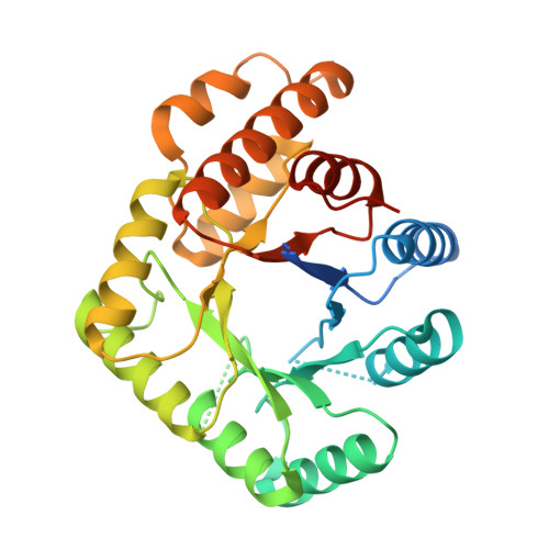
Structural Perturbations in the Ala -> Val Polymorphism of Methylenetetrahydrofolate Reductase: How Binding of Folates May Protect against Inactivation
Pejchal, R., Campbell, E., Guenther, B.D., Lennon, B.W., Matthews, R.G., Ludwig, M.L.(2006) Biochemistry 45: 4808-4818
- PubMed: 16605249
- DOI: https://doi.org/10.1021/bi052294c
- Primary Citation of Related Structures:
2FMN, 2FMO - PubMed Abstract:
In human methylenetetrahydrofolate reductase (MTHFR) the Ala222Val (677C-->T) polymorphism encodes a heat-labile gene product that is associated with elevated levels of homocysteine and possibly with risk for cardiovascular disease. Generation of the equivalent Ala to Val mutation in Escherichia coli MTHFR, which is 30% identical to the catalytic domain of the human enzyme, creates a protein with enhanced thermolability. In both human and E. coli MTHFR, the A --> V mutation increases the rate of dissociation of FAD, and in both enzymes, loss of FAD is linked to changes in quaternary structure [Yamada, K., Chen, Z., Rozen, R., and Matthews, R. G. (2001) Proc. Natl. Acad. Sci. U.S.A. 98, 14853-14858; Guenther, B. D., Sheppard, C. A., Tran, P., Rozen, R., Matthews, R. G., and Ludwig, M. L. (1999) Nat. Struct. Biol. 6, 359-365]. Folates have been shown to protect both human and bacterial enzymes from loss of FAD. Despite its effect on affinity for FAD, the A --> V mutation is located at the bottom of the (betaalpha)(8) barrel of the catalytic domain in a position that does not contact the bound FAD prosthetic group. Here we report the structures of the Ala177Val mutant of E. coli MTHFR and of its complex with the 5,10-dideazafolate analogue, LY309887, and suggest mechanisms by which the mutation may perturb FAD binding. Helix alpha5, which immediately precedes the loop bearing the mutation, carries several residues that interact with FAD, including Asn168, Arg171, and Lys172. In the structures of the mutant enzyme this helix is displaced, perturbing protein-FAD interactions. In the complex with LY309887, the pterin-like ring of the analogue stacks against the si face of the flavin and is secured by hydrogen bonds to residues Gln183 and Asp120 that adjoin this face. The direct interactions of bound folate with the cofactor provide one mechanism for linkage between binding of FAD and folate binding that could account in part for the protective action of folates. Conformation changes induced by folate binding may also suppress dissociation of FAD.
Organizational Affiliation:
Department of Biological Chemistry, the Biophysics Research Division, and the Life Sciences Institute, The University of Michigan, Ann Arbor, Michigan 48109, USA.


