2NZD

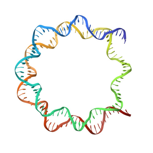
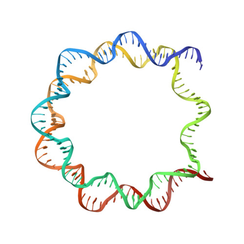
Nucleosome core particle containing 145 bp of DNA


Experimental Data Snapshot

  • Method: X-RAY DIFFRACTION
  • Resolution: 2.65 Å
  • R-Value Free: 0.283 
  • R-Value Work: 0.233 
  • R-Value Observed: 0.234 

Starting Model: experimental
View more details

wwPDB Validation   3D Report Full Report


This is version 1.3 of the entry. See complete history


Literature

DNA stretching and extreme kinking in the nucleosome core

Ong, M.S.Richmond, T.J.Davey, C.A.

(2007) J Mol Biol 368: 1067-1074

  • DOI: https://doi.org/10.1016/j.jmb.2007.02.062
  • Primary Citation of Related Structures:  
    2NZD

  • PubMed Abstract: 

    DNA stretching in chromatin may facilitate its compaction and influence site recognition by nuclear factors. In vivo, stretching has been estimated to occur at the equivalent of one to two base-pairs (bp) per nucleosome. We have determined the crystal structure of a nucleosome core particle containing 145 bp of DNA (NCP145). Compared to the structure with 147 bp, the NCP145 displays two incidences of stretching one to two double-helical turns from the particle dyad axis. The stretching illustrates clearly a mechanism for shifting DNA position by displacement of a single base-pair while maintaining nearly identical histone-DNA interactions. Increased DNA twist localized to a short section between adjacent histone-DNA binding sites advances the rotational setting, while a translational component involves DNA kinking at a flanking region that initiates elongation by unstacking bases. Furthermore, one stretched region of the NCP145 displays an extraordinary 55 degrees kink into the minor groove situated 1.5 double-helical turns from the particle dyad axis, a hot spot for gene insertion by HIV-integrase, which prefers highly distorted substrate. This suggests that nucleosome position and context within chromatin could promote extreme DNA kinking that may influence genomic processes.


  • Organizational Affiliation

    Division of Structural and Computational Biology, School of Biological Sciences, Nanyang Technological University, 60 Nanyang Drive, Singapore 637551, Singapore.


Macromolecules

Find similar proteins by:  (by identity cutoff)  |  3D Structure
Entity ID: 3
MoleculeChains Sequence LengthOrganismDetailsImage
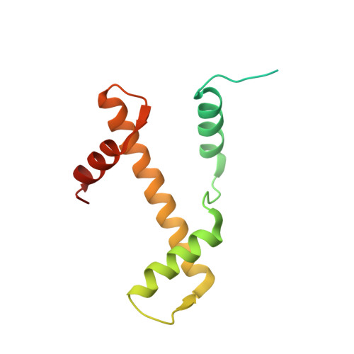
Histone H3C [auth A],
G [auth E]
135Xenopus laevisMutation(s): 2 
UniProt
Find proteins for P84233 (Xenopus laevis)
Explore P84233 
Go to UniProtKB:  P84233
Entity Groups  
Sequence Clusters30% Identity50% Identity70% Identity90% Identity95% Identity100% Identity
UniProt GroupP84233
Sequence Annotations
Expand
  • Reference Sequence
Find similar proteins by:  (by identity cutoff)  |  3D Structure
Entity ID: 4
MoleculeChains Sequence LengthOrganismDetailsImage
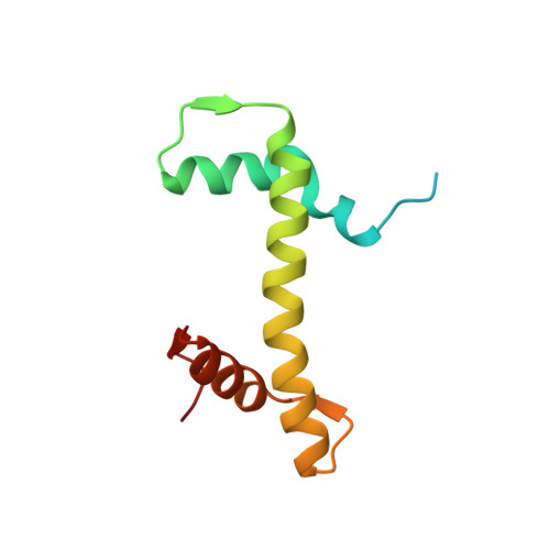
Histone H4D [auth B],
H [auth F]
102Xenopus laevisMutation(s): 0 
UniProt
Find proteins for P62799 (Xenopus laevis)
Explore P62799 
Go to UniProtKB:  P62799
Entity Groups  
Sequence Clusters30% Identity50% Identity70% Identity90% Identity95% Identity100% Identity
UniProt GroupP62799
Sequence Annotations
Expand
  • Reference Sequence
Find similar proteins by:  (by identity cutoff)  |  3D Structure
Entity ID: 5
MoleculeChains Sequence LengthOrganismDetailsImage
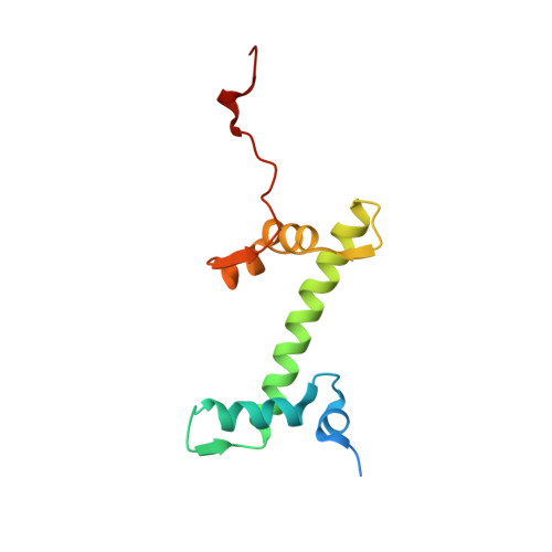
histone H2AE [auth C],
I [auth G]
119Xenopus laevisMutation(s): 0 
UniProt
Find proteins for P06897 (Xenopus laevis)
Explore P06897 
Go to UniProtKB:  P06897
Entity Groups  
Sequence Clusters30% Identity50% Identity70% Identity90% Identity95% Identity100% Identity
UniProt GroupP06897
Sequence Annotations
Expand
  • Reference Sequence
Find similar proteins by:  (by identity cutoff)  |  3D Structure
Entity ID: 6
MoleculeChains Sequence LengthOrganismDetailsImage
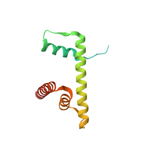
Histone H2BF [auth D],
J [auth H]
125Xenopus laevisMutation(s): 1 
UniProt
Find proteins for P02281 (Xenopus laevis)
Explore P02281 
Go to UniProtKB:  P02281
Entity Groups  
Sequence Clusters30% Identity50% Identity70% Identity90% Identity95% Identity100% Identity
UniProt GroupP02281
Sequence Annotations
Expand
  • Reference Sequence
Find similar nucleic acids by:  (by identity cutoff)  |  3D Structure
Entity ID: 1
MoleculeChains LengthOrganismImage
DNA (145-MER)A [auth I]145N/A
Sequence Annotations
Expand
  • Reference Sequence
Find similar nucleic acids by:  (by identity cutoff)  |  3D Structure
Entity ID: 2
MoleculeChains LengthOrganismImage
DNA (145-MER)B [auth J]145N/A
Sequence Annotations
Expand
  • Reference Sequence
Small Molecules
Ligands 1 Unique
IDChains Name / Formula / InChI Key2D Diagram3D Interactions
MN
Query on MN

Download Ideal Coordinates CCD File 
K [auth I]
L [auth I]
M [auth I]
N [auth I]
O [auth I]
K [auth I],
L [auth I],
M [auth I],
N [auth I],
O [auth I],
P [auth J],
Q [auth J],
R [auth J],
S [auth J],
T [auth J],
U [auth E]
MANGANESE (II) ION
Mn
WAEMQWOKJMHJLA-UHFFFAOYSA-N
Experimental Data & Validation

Experimental Data

  • Method: X-RAY DIFFRACTION
  • Resolution: 2.65 Å
  • R-Value Free: 0.283 
  • R-Value Work: 0.233 
  • R-Value Observed: 0.234 
  • Space Group: P 21 21 21
Unit Cell:
Length ( Å )Angle ( ˚ )
a = 105.617α = 90
b = 109.856β = 90
c = 181.217γ = 90
Software Package:
Software NamePurpose
SCALAdata scaling
REFMACrefinement
PDB_EXTRACTdata extraction
CrystalCleardata collection
MOSFLMdata reduction
CCP4data scaling
CNSphasing

Structure Validation

View Full Validation Report


Entry History 

Deposition Data

Revision History  (Full details and data files)

  • Version 1.0: 2007-04-10
    Type: Initial release
  • Version 1.1: 2008-05-01
    Changes: Version format compliance
  • Version 1.2: 2011-07-13
    Changes: Version format compliance
  • Version 1.3: 2023-08-30
    Changes: Data collection, Database references, Derived calculations, Refinement description