2OU7

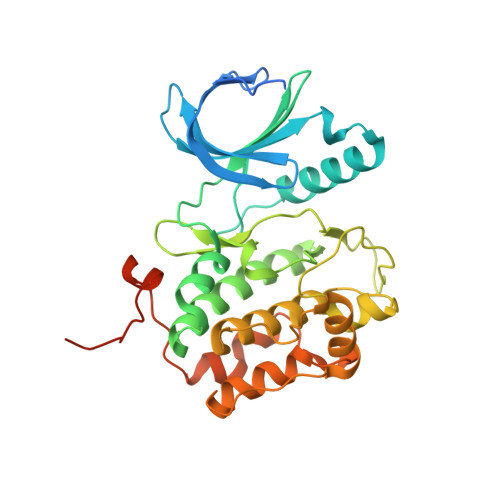
Structure of the Catalytic Domain of Human Polo-like Kinase 1


Experimental Data Snapshot

  • Method: X-RAY DIFFRACTION
  • Resolution: 2.40 Å
  • R-Value Free: 0.235 
  • R-Value Work: 0.203 
  • R-Value Observed: 0.205 

Starting Model: experimental
View more details

wwPDB Validation   3D Report Full Report


This is version 1.5 of the entry. See complete history


Literature

Structure of the catalytic domain of human polo-like kinase 1.

Kothe, M.Kohls, D.Low, S.Coli, R.Cheng, A.C.Jacques, S.L.Johnson, T.L.Lewis, C.Loh, C.Nonomiya, J.Sheils, A.L.Verdries, K.A.Wynn, T.A.Kuhn, C.Ding, Y.H.

(2007) Biochemistry 46: 5960-5971

  • DOI: https://doi.org/10.1021/bi602474j
  • Primary Citation of Related Structures:  
    2OU7, 2OWB

  • PubMed Abstract: 

    Polo-like kinase 1 (Plk1) is an attractive target for the development of anticancer agents due to its importance in regulating cell-cycle progression. Overexpression of Plk1 has been detected in a variety of cancers, and expression levels often correlate with poor prognosis. Despite high interest in Plk1-targeted therapeutics, there is currently no structure publicly available to guide structure-based drug design of specific inhibitors. We determined the crystal structures of the T210V mutant of the kinase domain of human Plk1 complexed with the nonhydrolyzable ATP analogue adenylylimidodiphosphate (AMPPNP) or the pyrrolo-pyrazole inhibitor PHA-680626 at 2.4 and 2.1 A resolution, respectively. Plk1 adopts the typical kinase domain fold and crystallized in a conformation resembling the active state of other kinases. Comparison of the kinetic parameters determined for the (unphosphorylated) wild-type enzyme, as well as the T210V and T210D mutants, shows that the mutations primarily affect the kcat of the reaction, with little change in the apparent Km for the protein or nucleotide substrates (kcat = 0.0094, 0.0376, and 0.0049 s-1 and Km(ATP) = 3.2, 4.0, and 3.0 microM for WT, T210D, and T210V, respectively). The structure highlights features of the active site that can be exploited to obtain Plk1-specific inhibitors with selectivity over other kinases and Plk isoforms. These include the presence of a phenylalanine at the bottom of the ATP pocket, combined with a cysteine (as opposed to the more commonly found leucine) in the roof of the binding site, a pocket created by Leu132 in the hinge region, and a cluster of positively charged residues in the solvent-exposed area outside of the adenine pocket adjacent to the hinge region.


  • Organizational Affiliation

    Pfizer Global Research and Development, Research Technology Center, 620 Memorial Drive, Cambridge, Massachusetts 02139, USA.


Macromolecules
Find similar proteins by:  (by identity cutoff)  |  3D Structure
Entity ID: 1
MoleculeChains Sequence LengthOrganismDetailsImage
Serine/threonine-protein kinase PLK1335Homo sapiensMutation(s): 1 
Gene Names: PLK1PLK
EC: 2.7.11.21
UniProt & NIH Common Fund Data Resources
Find proteins for P53350 (Homo sapiens)
Explore P53350 
Go to UniProtKB:  P53350
PHAROS:  P53350
GTEx:  ENSG00000166851 
Entity Groups  
Sequence Clusters30% Identity50% Identity70% Identity90% Identity95% Identity100% Identity
UniProt GroupP53350
Sequence Annotations
Expand
  • Reference Sequence
Small Molecules
Ligands 4 Unique
IDChains Name / Formula / InChI Key2D Diagram3D Interactions
ANP
Query on ANP

Download Ideal Coordinates CCD File 
G [auth A]PHOSPHOAMINOPHOSPHONIC ACID-ADENYLATE ESTER
C10 H17 N6 O12 P3
PVKSNHVPLWYQGJ-KQYNXXCUSA-N
ZN
Query on ZN

Download Ideal Coordinates CCD File 
B [auth A]ZINC ION
Zn
PTFCDOFLOPIGGS-UHFFFAOYSA-N
ACT
Query on ACT

Download Ideal Coordinates CCD File 
C [auth A],
D [auth A]
ACETATE ION
C2 H3 O2
QTBSBXVTEAMEQO-UHFFFAOYSA-M
MG
Query on MG

Download Ideal Coordinates CCD File 
E [auth A],
F [auth A]
MAGNESIUM ION
Mg
JLVVSXFLKOJNIY-UHFFFAOYSA-N
Binding Affinity Annotations 
IDSourceBinding Affinity
ANP PDBBind:  2OU7 IC50: 1.55e+5 (nM) from 1 assay(s)
Experimental Data & Validation

Experimental Data

  • Method: X-RAY DIFFRACTION
  • Resolution: 2.40 Å
  • R-Value Free: 0.235 
  • R-Value Work: 0.203 
  • R-Value Observed: 0.205 
  • Space Group: P 32 2 1
Unit Cell:
Length ( Å )Angle ( ˚ )
a = 65.958α = 90
b = 65.958β = 90
c = 154.045γ = 120
Software Package:
Software NamePurpose
REFMACrefinement
JDirectordata collection
HKL-2000data reduction
HKL-2000data scaling
MOLREPphasing

Structure Validation

View Full Validation Report


Entry History 

Deposition Data

Revision History  (Full details and data files)

  • Version 1.0: 2007-04-24
    Type: Initial release
  • Version 1.1: 2008-05-01
    Changes: Version format compliance
  • Version 1.2: 2011-07-13
    Changes: Derived calculations, Version format compliance
  • Version 1.3: 2017-10-18
    Changes: Refinement description
  • Version 1.4: 2021-10-20
    Changes: Database references, Derived calculations
  • Version 1.5: 2023-08-30
    Changes: Data collection, Refinement description