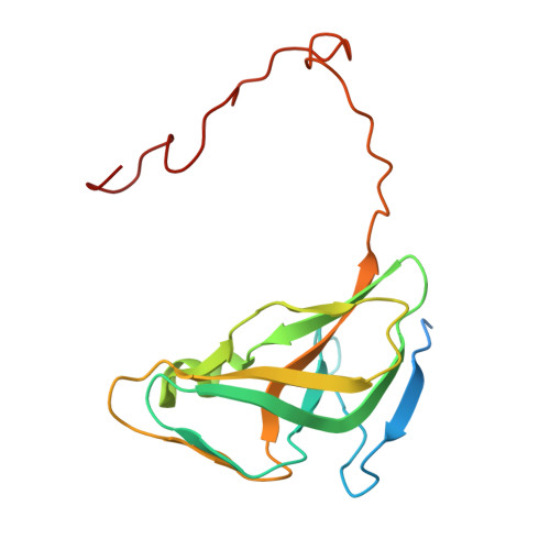
2PY4

Full length structure of the Mycobacterium tuberculosis dUTPase complexed with magnesium and alpha,beta-imido-dUTP.


Experimental Data Snapshot

  • Method: X-RAY DIFFRACTION
  • Resolution: 1.49 Å
  • R-Value Free: 0.170 
  • R-Value Work: 0.144 
  • R-Value Observed: 0.146 

Starting Model: experimental
View more details

wwPDB Validation   3D Report Full Report


Ligand Structure Quality Assessment 


This is version 1.3 of the entry. See complete history


Literature

Active site of mycobacterial dUTPase: structural characteristics and a built-in sensor.

Varga, B.Barabas, O.Takacs, E.Nagy, N.Nagy, P.Vertessy, B.G.

(2008) Biochem Biophys Res Commun 373: 8-13

  • DOI: https://doi.org/10.1016/j.bbrc.2008.05.130
  • Primary Citation of Related Structures:  
    2PY4

  • PubMed Abstract: 

    dUTPases are essential to eliminate dUTP for DNA integrity and provide dUMP for thymidylate biosynthesis. Mycobacterium tuberculosis apparently lacks any other thymidylate biosynthesis pathway, therefore dUTPase is a promising antituberculotic drug target. Crystal structure of the mycobacterial enzyme in complex with the isosteric substrate analog, alpha,beta-imido-dUTP and Mg(2+) at 1.5A resolution was determined that visualizes the full-length C-terminus, previously not localized. Interactions of a conserved motif important in catalysis, the Mycobacterium-specific five-residue-loop insert and C-terminal tetrapeptide could now be described in detail. Stacking of C-terminal histidine upon the uracil moiety prompted replacement with tryptophan. The resulting sensitive fluorescent sensor enables fast screening for binding of potential inhibitors to the active site. K(d) for alpha,beta-imido-dUTP binding to mycobacterial dUTPase is determined to be 10-fold less than for human dUTPase, which is to be considered in drug optimization. A robust continuous activity assay for kinetic screening is proposed.


  • Organizational Affiliation

    Laboratory of Genome Metabolism and Repair, Institute of Enzymology, Hungarian Academy of Sciences, Karolina ut 29, H-1113 Budapest, Hungary.


Macromolecules
Find similar proteins by:  (by identity cutoff)  |  3D Structure
Entity ID: 1
MoleculeChains Sequence LengthOrganismDetailsImage
Deoxyuridine 5'-triphosphate nucleotidohydrolase174Mycobacterium tuberculosisMutation(s): 0 
Gene Names: dut
EC: 3.6.1.23
UniProt
Find proteins for P9WNS5 (Mycobacterium tuberculosis (strain ATCC 25618 / H37Rv))
Explore P9WNS5 
Go to UniProtKB:  P9WNS5
Entity Groups  
Sequence Clusters30% Identity50% Identity70% Identity90% Identity95% Identity100% Identity
UniProt GroupP9WNS5
Sequence Annotations
Expand
  • Reference Sequence
Small Molecules
Binding Affinity Annotations 
IDSourceBinding Affinity
DUP PDBBind:  2PY4 Kd: 200 (nM) from 1 assay(s)
Experimental Data & Validation

Experimental Data

  • Method: X-RAY DIFFRACTION
  • Resolution: 1.49 Å
  • R-Value Free: 0.170 
  • R-Value Work: 0.144 
  • R-Value Observed: 0.146 
  • Space Group: P 63
Unit Cell:
Length ( Å )Angle ( ˚ )
a = 54.591α = 90
b = 54.591β = 90
c = 83.051γ = 120
Software Package:
Software NamePurpose
REFMACrefinement
STARTdata collection
XDSdata reduction
XSCALEdata scaling
MOLREPphasing

Structure Validation

View Full Validation Report


Ligand Structure Quality Assessment 


Entry History 

Deposition Data

Revision History  (Full details and data files)

  • Version 1.0: 2007-05-22
    Type: Initial release
  • Version 1.1: 2008-05-01
    Changes: Version format compliance
  • Version 1.2: 2011-07-13
    Changes: Advisory, Derived calculations, Version format compliance
  • Version 1.3: 2023-08-30
    Changes: Data collection, Database references, Derived calculations, Refinement description