2ZZX

Crystal Structure of a Periplasmic Substrate Binding Protein in Complex with Lactate


Experimental Data Snapshot

  • Method: X-RAY DIFFRACTION
  • Resolution: 1.75 Å
  • R-Value Free: 0.207 
  • R-Value Work: 0.177 
  • R-Value Observed: 0.177 

Starting Model: experimental
View more details

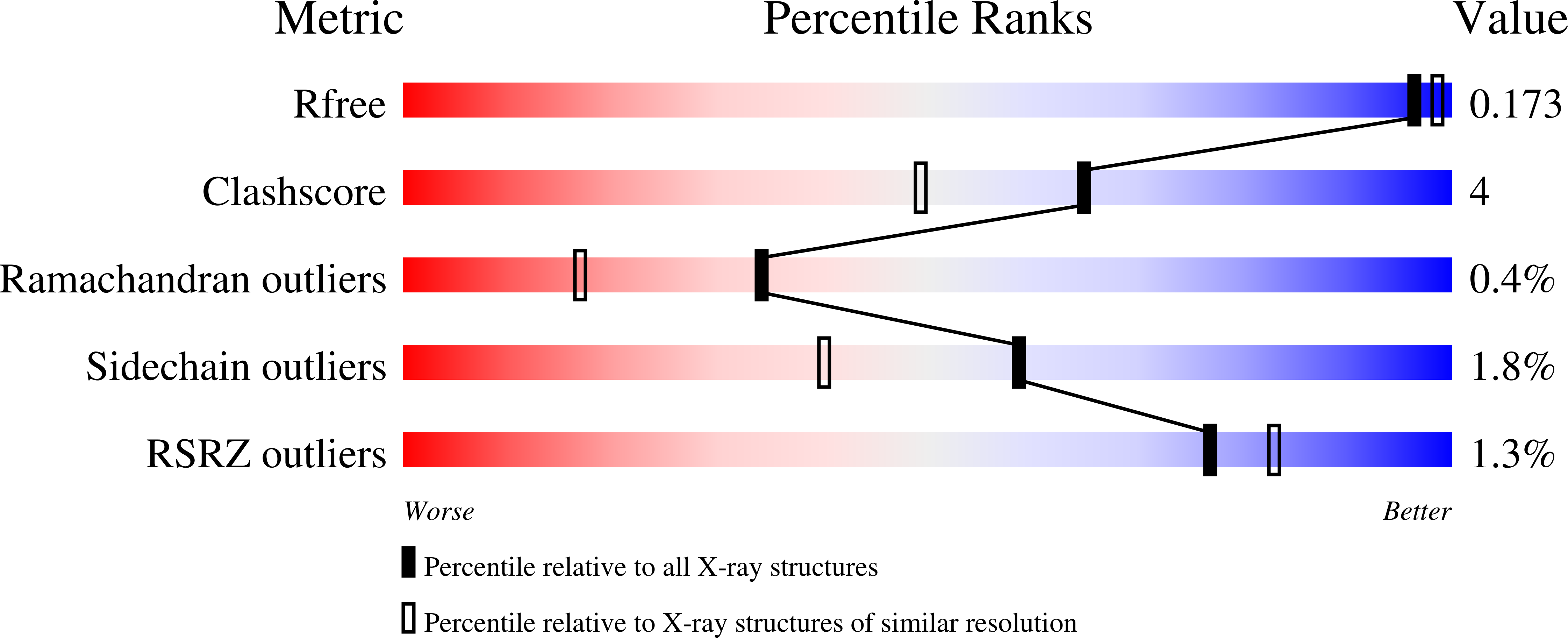
wwPDB Validation   3D Report Full Report


This is version 2.0 of the entry. See complete history


Literature

Crystal structure of a periplasmic substrate-binding protein in complex with calcium lactate

Akiyama, N.Takeda, K.Miki, K.

(2009) J Mol Biol 392: 559-565

  • DOI: https://doi.org/10.1016/j.jmb.2009.07.043
  • Primary Citation of Related Structures:  
    2ZZV, 2ZZW, 2ZZX

  • PubMed Abstract: 

    Lactate is utilized in many biological processes, and its transport across biological membranes is mediated with various types of transporters. Here, we report the crystal structures of a lactate-binding protein of a TRAP (tripartite ATP-independent periplasmic) secondary transporter from Thermus thermophilus HB8. The folding of the protein is typical for a type II periplasmic solute-binding protein and forms a dimer in a back-to-back manner. One molecule of l-lactate is clearly identified in a cleft of the protein as a complex with a calcium ion. Detailed crystallographic and biochemical analyses revealed that the calcium ion can be removed from the protein and replaced with other divalent cations. This characterization of the structure of a protein binding with calcium lactate makes a significant contribution to our understanding of the mechanisms by which calcium and lactate are accommodated in cells.


  • Organizational Affiliation

    Department of Chemistry, Kyoto University, Sakyo-ku, Japan.


Macromolecules
Find similar proteins by:  (by identity cutoff)  |  3D Structure
Entity ID: 1
MoleculeChains Sequence LengthOrganismDetailsImage
ABC transporter, solute-binding protein
A, B, C, D
361Thermus thermophilus HB8Mutation(s): 0 
Gene Names: TTHA0766
UniProt
Find proteins for Q5SK82 (Thermus thermophilus (strain ATCC 27634 / DSM 579 / HB8))
Explore Q5SK82 
Go to UniProtKB:  Q5SK82
Entity Groups  
Sequence Clusters30% Identity50% Identity70% Identity90% Identity95% Identity100% Identity
UniProt GroupQ5SK82
Sequence Annotations
Expand
  • Reference Sequence
Small Molecules
Ligands 4 Unique
IDChains Name / Formula / InChI Key2D Diagram3D Interactions
PO4
Query on PO4

Download Ideal Coordinates CCD File 
F [auth A],
I [auth B]
PHOSPHATE ION
O4 P
NBIIXXVUZAFLBC-UHFFFAOYSA-K
GOL
Query on GOL

Download Ideal Coordinates CCD File 
G [auth A],
M [auth D]
GLYCEROL
C3 H8 O3
PEDCQBHIVMGVHV-UHFFFAOYSA-N
LAC
Query on LAC

Download Ideal Coordinates CCD File 
E [auth A],
H [auth B],
J [auth C],
L [auth D]
LACTIC ACID
C3 H6 O3
JVTAAEKCZFNVCJ-UWTATZPHSA-N
CA
Query on CA

Download Ideal Coordinates CCD File 
K [auth C]CALCIUM ION
Ca
BHPQYMZQTOCNFJ-UHFFFAOYSA-N
Experimental Data & Validation

Experimental Data

  • Method: X-RAY DIFFRACTION
  • Resolution: 1.75 Å
  • R-Value Free: 0.207 
  • R-Value Work: 0.177 
  • R-Value Observed: 0.177 
  • Space Group: C 1 2 1
Unit Cell:
Length ( Å )Angle ( ˚ )
a = 162.792α = 90
b = 103.501β = 101.86
c = 90.76γ = 90
Software Package:
Software NamePurpose
CNSrefinement
HKL-2000data collection
HKL-2000data reduction
HKL-2000data scaling
MOLREPphasing

Structure Validation

View Full Validation Report


Entry History 

Deposition Data

Revision History  (Full details and data files)

  • Version 1.0: 2009-08-11
    Type: Initial release
  • Version 1.1: 2011-07-13
    Changes: Non-polymer description, Version format compliance
  • Version 1.2: 2023-11-01
    Changes: Data collection, Database references, Derived calculations, Refinement description
  • Version 2.0: 2023-11-15
    Changes: Atomic model, Data collection, Derived calculations