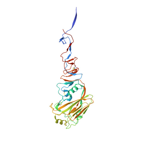
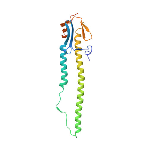
3AL4

Crystal structure of the swine-origin A (H1N1)-2009 influenza A virus hemagglutinin (HA) reveals similar antigenicity to that of the 1918 pandemic virus


Experimental Data Snapshot

  • Method: X-RAY DIFFRACTION
  • Resolution: 2.87 Å
  • R-Value Free: 0.270 
  • R-Value Work: 0.246 
  • R-Value Observed: 0.247 

Starting Model: experimental
View more details

wwPDB Validation   3D Report Full Report


Ligand Structure Quality Assessment 


This is version 2.2 of the entry. See complete history


Literature

Crystal structure of the swine-origin A (H1N1)-2009 influenza A virus hemagglutinin (HA) reveals similar antigenicity to that of the 1918 pandemic virus

Zhang, W.Qi, J.Shi, Y.Li, Q.Gao, F.Sun, Y.Lu, X.Lu, Q.Vavricka, C.J.Liu, D.Yan, J.Gao, G.F.

(2010) Protein Cell 1: 459-467

  • DOI: https://doi.org/10.1007/s13238-010-0059-1
  • Primary Citation of Related Structures:  
    3AL4

  • PubMed Abstract: 

    Influenza virus is the causative agent of the seasonal and occasional pandemic flu. The current H1N1 influenza pandemic, announced by the WHO in June 2009, is highly contagious and responsible for global economic losses and fatalities. Although the H1N1 gene segments have three origins in terms of host species, the virus has been named swine-origin influenza virus (S-OIV) due to a predominant swine origin. 2009 S-OIV has been shown to highly resemble the 1918 pandemic virus in many aspects. Hemagglutinin is responsible for the host range and receptor binding of the virus and is therefore a primary indicator for the potential of infection. Primary sequence analysis of the 2009 S-OIV hemagglutinin (HA) reveals its closest relationship to that of the 1918 pandemic influenza virus, however, analysis at the structural level is necessary to critically assess the functional significance. In this report, we report the crystal structure of soluble hemagglutinin H1 (09H1) at 2.9 Å, illustrating that the 09H1 is very similar to the 1918 pandemic HA (18H1) in overall structure and the structural modules, including the five defined antiboby (Ab)-binding epitopes. Our results provide an explanation as to why sera from the survivors of the 1918 pandemics can neutralize the 2009 S-OIV, and people born around the 1918 are resistant to the current pandemic, yet younger generations are more susceptible to the 2009 pandemic.


  • Organizational Affiliation

    CAS Key Laboratory of Pathogenic Microbiology and Immunology, Institute of Microbiology, Chinese Academy of Sciences, Beijing 100101, China.


Macromolecules
Find similar proteins by:  (by identity cutoff)  |  3D Structure
Entity ID: 1
MoleculeChains Sequence LengthOrganismDetailsImage
Hemagglutinin
A, C, E, G, I
A, C, E, G, I, K
333Influenza A virus (A/California/04/2009(H1N1))Mutation(s): 0 
Gene Names: HEMAGGLUTININ
UniProt
Find proteins for C3W5S1 (Influenza A virus (strain swl A/California/04/2009 H1N1))
Explore C3W5S1 
Go to UniProtKB:  C3W5S1
Entity Groups  
Sequence Clusters30% Identity50% Identity70% Identity90% Identity95% Identity100% Identity
UniProt GroupC3W5S1
Glycosylation
Glycosylation Sites: 4
Sequence Annotations
Expand
  • Reference Sequence
Find similar proteins by:  (by identity cutoff)  |  3D Structure
Entity ID: 2
MoleculeChains Sequence LengthOrganismDetailsImage
Hemagglutinin
B, D, F, H, J
B, D, F, H, J, L
181Influenza A virus (A/California/04/2009(H1N1))Mutation(s): 0 
Gene Names: HEMAGGLUTININ
UniProt
Find proteins for C3W5S1 (Influenza A virus (strain swl A/California/04/2009 H1N1))
Explore C3W5S1 
Go to UniProtKB:  C3W5S1
Entity Groups  
Sequence Clusters30% Identity50% Identity70% Identity90% Identity95% Identity100% Identity
UniProt GroupC3W5S1
Glycosylation
Glycosylation Sites: 1
Sequence Annotations
Expand
  • Reference Sequence
Oligosaccharides

Help

Entity ID: 3
MoleculeChains Length2D Diagram Glycosylation3D Interactions
2-acetamido-2-deoxy-beta-D-glucopyranose-(1-4)-2-acetamido-2-deoxy-beta-D-glucopyranose
M, N, P, Q, S
2N-Glycosylation
Glycosylation Resources
GlyTouCan:  G42666HT
GlyCosmos:  G42666HT
GlyGen:  G42666HT
Entity ID: 4
MoleculeChains Length2D Diagram Glycosylation3D Interactions
beta-D-mannopyranose-(1-4)-2-acetamido-2-deoxy-beta-D-glucopyranose-(1-4)-2-acetamido-2-deoxy-beta-D-glucopyranose
O, R
3N-Glycosylation
Glycosylation Resources
GlyTouCan:  G15407YE
GlyCosmos:  G15407YE
GlyGen:  G15407YE
Small Molecules
Ligands 1 Unique
IDChains Name / Formula / InChI Key2D Diagram3D Interactions
NAG
Query on NAG

Download Ideal Coordinates CCD File 
AA [auth F]
BA [auth G]
CA [auth G]
DA [auth G]
EA [auth K]
AA [auth F],
BA [auth G],
CA [auth G],
DA [auth G],
EA [auth K],
FA [auth K],
GA [auth K],
HA [auth K],
IA [auth L],
T [auth A],
U [auth A],
V [auth C],
W [auth C],
X [auth E],
Y [auth E],
Z [auth E]
2-acetamido-2-deoxy-beta-D-glucopyranose
C8 H15 N O6
OVRNDRQMDRJTHS-FMDGEEDCSA-N
Experimental Data & Validation

Experimental Data

  • Method: X-RAY DIFFRACTION
  • Resolution: 2.87 Å
  • R-Value Free: 0.270 
  • R-Value Work: 0.246 
  • R-Value Observed: 0.247 
  • Space Group: P 1
Unit Cell:
Length ( Å )Angle ( ˚ )
a = 66.02α = 62.31
b = 115.19β = 77.94
c = 114.985γ = 81.05
Software Package:
Software NamePurpose
MAR345dtbdata collection
PHASERphasing
PHENIXrefinement
HKL-2000data reduction
SCALEPACKdata scaling

Structure Validation

View Full Validation Report


Ligand Structure Quality Assessment 


Entry History 

Deposition Data

Revision History  (Full details and data files)

  • Version 1.0: 2010-08-04
    Type: Initial release
  • Version 1.1: 2011-07-13
    Changes: Version format compliance
  • Version 1.2: 2012-06-27
    Changes: Database references
  • Version 2.0: 2020-07-29
    Type: Remediation
    Reason: Carbohydrate remediation
    Changes: Advisory, Atomic model, Data collection, Database references, Derived calculations, Structure summary
  • Version 2.1: 2023-11-01
    Changes: Data collection, Database references, Refinement description, Structure summary
  • Version 2.2: 2024-10-23
    Changes: Structure summary