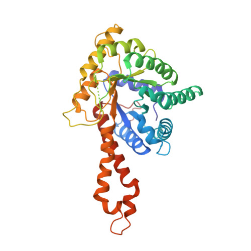
3EKZ

Structural Characterization of tetrameric Mycobacterium tuberculosis fructose 1,6-bisphosphate aldolase - substrate binding and catalysis mechanism of a class IIa bacterial aldolase


Experimental Data Snapshot

  • Method: X-RAY DIFFRACTION
  • Resolution: 2.07 Å
  • R-Value Free: 0.180 
  • R-Value Work: 0.158 
  • R-Value Observed: 0.159 

Starting Model: experimental
View more details

wwPDB Validation   3D Report Full Report


Ligand Structure Quality Assessment 


This is version 1.3 of the entry. See complete history


Literature

Structural basis for catalysis of a tetrameric class IIa fructose 1,6-bisphosphate aldolase from Mycobacterium tuberculosis

Pegan, S.D.Rukseree, K.Franzblau, S.G.Mesecar, A.D.

(2009) J Mol Biol 386: 1038-1053

  • DOI: https://doi.org/10.1016/j.jmb.2009.01.003
  • Primary Citation of Related Structures:  
    3EKL, 3EKZ, 3ELF

  • PubMed Abstract: 

    Mycobacterium tuberculosis, the causative agent of tuberculosis (TB), currently infects one-third of the world's population in its latent form. The emergence of multidrug-resistant and extensive drug-resistant strains has highlighted the need for new pharmacological targets within M. tuberculosis. The class IIa fructose 1,6-bisphosphate aldolase (FBA) enzyme from M. tuberculosis (MtFBA) has been proposed as one such target since it is upregulated in latent TB. Since the structure of MtFBA has not been determined and there is little information available on its reaction mechanism, we sought to determine the X-ray structure of MtFBA in complex with its substrates. By lowering the pH of the enzyme in the crystalline state, we were able to determine a series of high-resolution X-ray structures of MtFBA bound to dihydroxyacetone phosphate, glyceraldehyde 3-phosphate, and fructose 1,6-bisphosphate at 1.5, 2.1, and 1.3 A, respectively. Through these structures, it was discovered that MtFBA belongs to a novel tetrameric class of type IIa FBAs. The molecular details at the interface of the tetramer revealed important information for better predictability of the quaternary structures among the FBAs based on their primary sequences. These X-ray structures also provide interesting and new details on the reaction mechanism of class II FBAs. Substrates and products were observed in geometries poised for catalysis; in addition, unexpectedly, the hydroxyl-enolate intermediate of dihydroxyacetone phosphate was also captured and resolved structurally. These concise new details offer a better understanding of the reaction mechanisms for FBAs in general and provide a structural basis for inhibitor design efforts aimed at this class of enzymes.


  • Organizational Affiliation

    Department of Medicinal Chemistry and Pharmacognosy, University of Illinois at Chicago, Chicago, IL 60607, USA.


Macromolecules
Find similar proteins by:  (by identity cutoff)  |  3D Structure
Entity ID: 1
MoleculeChains Sequence LengthOrganismDetailsImage
Fructose-bisphosphate aldolase349Mycobacterium tuberculosisMutation(s): 0 
Gene Names: fbaMT0379MTCY13E10.25cRv0363c
EC: 4.1.2.13
UniProt
Find proteins for P9WQA3 (Mycobacterium tuberculosis (strain ATCC 25618 / H37Rv))
Explore P9WQA3 
Go to UniProtKB:  P9WQA3
Entity Groups  
Sequence Clusters30% Identity50% Identity70% Identity90% Identity95% Identity100% Identity
UniProt GroupP9WQA3
Sequence Annotations
Expand
  • Reference Sequence
Small Molecules
Ligands 4 Unique
IDChains Name / Formula / InChI Key2D Diagram3D Interactions
G3P
Query on G3P

Download Ideal Coordinates CCD File 
D [auth A]SN-GLYCEROL-3-PHOSPHATE
C3 H9 O6 P
AWUCVROLDVIAJX-GSVOUGTGSA-N
13P
Query on 13P

Download Ideal Coordinates CCD File 
B [auth A],
C [auth A]
1,3-DIHYDROXYACETONEPHOSPHATE
C3 H7 O6 P
GNGACRATGGDKBX-UHFFFAOYSA-N
ZN
Query on ZN

Download Ideal Coordinates CCD File 
F [auth A],
G [auth A]
ZINC ION
Zn
PTFCDOFLOPIGGS-UHFFFAOYSA-N
NA
Query on NA

Download Ideal Coordinates CCD File 
E [auth A]SODIUM ION
Na
FKNQFGJONOIPTF-UHFFFAOYSA-N
Experimental Data & Validation

Experimental Data

  • Method: X-RAY DIFFRACTION
  • Resolution: 2.07 Å
  • R-Value Free: 0.180 
  • R-Value Work: 0.158 
  • R-Value Observed: 0.159 
  • Space Group: I 2 2 2
Unit Cell:
Length ( Å )Angle ( ˚ )
a = 61.22α = 90
b = 120.486β = 90
c = 164.327γ = 90
Software Package:
Software NamePurpose
CrystalCleardata collection
PHASERphasing
REFMACrefinement
HKL-2000data reduction
HKL-2000data scaling

Structure Validation

View Full Validation Report


Ligand Structure Quality Assessment 


Entry History 

Deposition Data

Revision History  (Full details and data files)

  • Version 1.0: 2009-02-10
    Type: Initial release
  • Version 1.1: 2011-07-13
    Changes: Version format compliance
  • Version 1.2: 2018-04-04
    Changes: Data collection
  • Version 1.3: 2023-08-30
    Changes: Data collection, Database references, Derived calculations, Refinement description