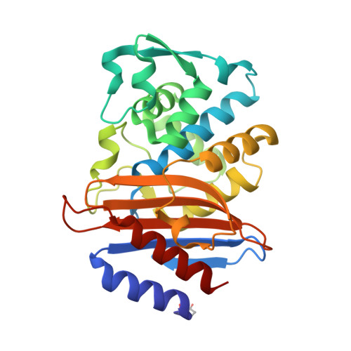
3G2Y

CTX-M-9 class A beta-lactamase complexed with compound 1 (GF4)


Experimental Data Snapshot

  • Method: X-RAY DIFFRACTION
  • Resolution: 1.31 Å
  • R-Value Free: 0.188 
  • R-Value Work: 0.161 
  • R-Value Observed: 0.162 

Starting Model: experimental
View more details

wwPDB Validation   3D Report Full Report


Ligand Structure Quality Assessment 


This is version 2.2 of the entry. See complete history


Literature

Molecular docking and ligand specificity in fragment-based inhibitor discovery

Chen, Y.Shoichet, B.K.

(2009) Nat Chem Biol 5: 358-364

  • DOI: https://doi.org/10.1038/nchembio.155
  • Primary Citation of Related Structures:  
    3G2Y, 3G2Z, 3G30, 3G31, 3G32, 3G34, 3G35

  • PubMed Abstract: 

    Fragment screens have successfully identified new scaffolds in drug discovery, often with relatively high hit rates (5%) using small screening libraries (1,000-10,000 compounds). This raises two questions: would other noteworthy chemotypes be found were one to screen all commercially available fragments (>300,000), and does the success rate imply low specificity of fragments? We used molecular docking to screen large libraries of fragments against CTX-M beta-lactamase. We identified ten millimolar-range inhibitors from the 69 compounds tested. The docking poses corresponded closely to the crystallographic structures subsequently determined. Notably, these initial low-affinity hits showed little specificity between CTX-M and an unrelated beta-lactamase, AmpC, which is unusual among beta-lactamase inhibitors. This is consistent with the idea that the high hit rates among fragments correlate to a low initial specificity. As the inhibitors were progressed, both specificity and affinity rose together, yielding to our knowledge the first micromolar-range noncovalent inhibitors against a class A beta-lactamase.


  • Organizational Affiliation

    Department of Pharmaceutical Chemistry, University of California, San Francisco, California, USA.


Macromolecules
Find similar proteins by:  (by identity cutoff)  |  3D Structure
Entity ID: 1
MoleculeChains Sequence LengthOrganismDetailsImage
Beta-lactamase CTX-M-9a
A, B
263Escherichia coliMutation(s): 0 
Gene Names: blaCTX-M-9blaCTX-M-9aCTX-M
EC: 3.5.2.6
UniProt
Find proteins for Q9L5C8 (Escherichia coli)
Explore Q9L5C8 
Go to UniProtKB:  Q9L5C8
Entity Groups  
Sequence Clusters30% Identity50% Identity70% Identity90% Identity95% Identity100% Identity
UniProt GroupQ9L5C8
Sequence Annotations
Expand
  • Reference Sequence
Small Molecules
Modified Residues  1 Unique
IDChains TypeFormula2D DiagramParent
PCA
Query on PCA
A, B
L-PEPTIDE LINKINGC5 H7 N O3GLN
Binding Affinity Annotations 
IDSourceBinding Affinity
GF4 PDBBind:  3G2Y Ki: 1.00e+7 (nM) from 1 assay(s)
Experimental Data & Validation

Experimental Data

  • Method: X-RAY DIFFRACTION
  • Resolution: 1.31 Å
  • R-Value Free: 0.188 
  • R-Value Work: 0.161 
  • R-Value Observed: 0.162 
  • Space Group: P 1 21 1
Unit Cell:
Length ( Å )Angle ( ˚ )
a = 45.154α = 90
b = 106.877β = 101.91
c = 47.703γ = 90
Software Package:
Software NamePurpose
DENZOdata reduction
SCALEPACKdata scaling
REFMACrefinement
PDB_EXTRACTdata extraction
HKL-2000data collection
HKL-2000data reduction
HKL-2000data scaling
REFMACphasing

Structure Validation

View Full Validation Report


Ligand Structure Quality Assessment 


Entry History 

Deposition Data

Revision History  (Full details and data files)

  • Version 1.0: 2009-03-24
    Type: Initial release
  • Version 1.1: 2011-07-13
    Changes: Version format compliance
  • Version 1.2: 2017-11-01
    Changes: Refinement description
  • Version 2.0: 2019-12-25
    Changes: Atomic model, Database references, Derived calculations, Polymer sequence
  • Version 2.1: 2023-09-06
    Changes: Data collection, Database references, Derived calculations, Refinement description
  • Version 2.2: 2024-11-06
    Changes: Structure summary