How to Replace the Residual Solvation Shell of Polar Active Site Residues to Achieve Nanomolar Inhibition of tRNA-Guanine Transglycosylase
Ritschel, T., Kohler, P.C., Neudert, G., Heine, A., Diederich, F., Klebe, G.(2009) ChemMedChem 4: 2012-2023
- PubMed: 19894214
- DOI: https://doi.org/10.1002/cmdc.200900343
- Primary Citation of Related Structures:
3EOS, 3EOU, 3GC4, 3GC5, 3GE7 - PubMed Abstract:
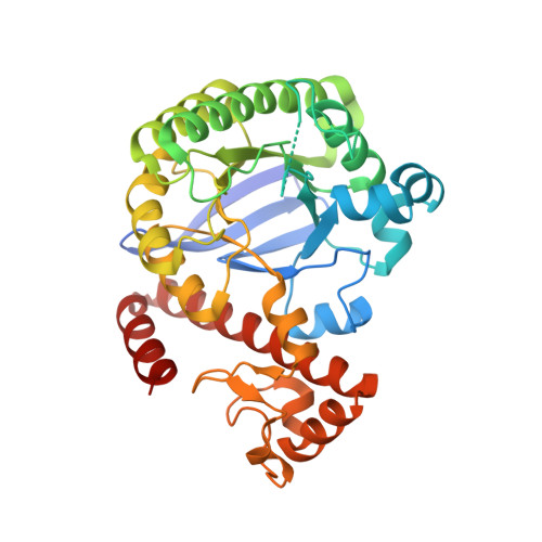
In a computational and structural study, we investigated a series of 4-substituted lin-benzoguanines that are potent inhibitors of tRNA-guanine transglycosylase (TGT), a putative target for the treatment of shigellosis. At first glance, it appears self-evident that the placement of a positively charged ligand functional group between the carboxylate groups of two adjacent aspartate residues in the glycosylase catalytic center leads to enhanced ligand binding. The concomitant displacement of water molecules that partially solvate the aspartates prior to ligand binding appears to result as a consequence of this. However, the case study presented herein shows that this premise is much too superficial. Placement of a likely positively charged amino group at such a pivotal position, interfering with the residual water solvation shell, is at best cost-neutral compared with the unsubstituted parent ligand not conflicting with the residual water shell. A ligand that orients a hydroxy group in this position shows even decreased binding. Based on the cost-neutral placement of the amino functionality, hydrophobic side chains can now be further attached to fill, with increasing potency, a small hydrophobic pocket remote to the aspartates. Any attempts to cross the pivotal position between both aspartates with nonpolar scaffolds reveals only decreased binding, even though the waters of the residual solvation shell are successfully repelled. This surprising observation fostered a detailed analysis of the role of water molecules involved in the residual solvation of polar active site residues. Their geometry and putative replacement in the binding pocket of TGT has been studied by a comparative database analysis, computational active site mapping, and a series of crystal structure analyses. Furthermore, conformational preferences of attached hydrophobic moieties explain their contribution to a gradual increase in binding affinity.
Organizational Affiliation:
Institut für Pharmazeutische Chemie, Philipps-Universität Marburg, Marbacher Weg 6, 35032 Marburg, Germany.


