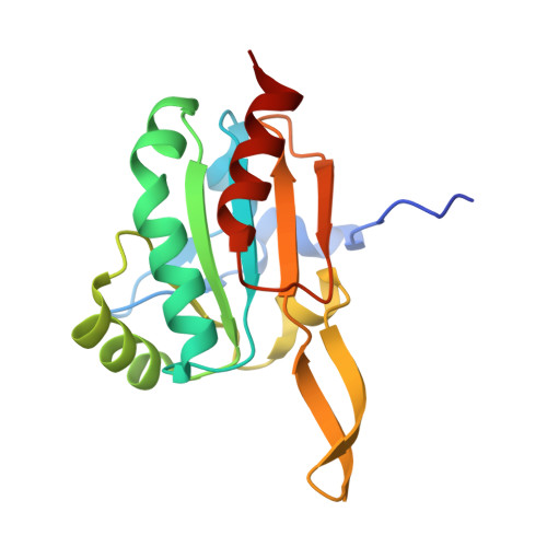
3GKN

Insights into the Alkyl Peroxide Reduction Activity of Xanthomonas campestris Bacterioferritin Comigratory Protein from the Trapped Intermediate/Ligand Complex Structures


Experimental Data Snapshot

  • Method: X-RAY DIFFRACTION
  • Resolution: 1.47 Å
  • R-Value Free: 0.211 
  • R-Value Work: 0.198 
  • R-Value Observed: 0.198 

Starting Model: experimental
View more details

wwPDB Validation   3D Report Full Report


Ligand Structure Quality Assessment 


This is version 1.5 of the entry. See complete history


Literature

Insights into the alkyl peroxide reduction pathway of Xanthomonas campestris bacterioferritin comigratory protein from the trapped intermediate-ligand complex structures

Liao, S.-J.Yang, C.-Y.Chin, K.-H.Wang, A.H.-J.Chou, S.-H.

(2009) J Mol Biol 390: 951-966

  • DOI: https://doi.org/10.1016/j.jmb.2009.05.030
  • Primary Citation of Related Structures:  
    3GKK, 3GKM, 3GKN

  • PubMed Abstract: 

    Considerable insights into the oxidoreduction activity of the Xanthomonas campestris bacterioferritin comigratory protein (XcBCP) have been obtained from trapped intermediate/ligand complex structures determined by X-ray crystallography. Multiple sequence alignment and enzyme assay indicate that XcBCP belongs to a subfamily of atypical 2-Cys peroxiredoxins (Prxs), containing a strictly conserved peroxidatic cysteine (C(P)48) and an unconserved resolving cysteine (C(R)84). Crystals at different states, i.e. Free_SH state, Intra_SS state, and Inter_SS state, were obtained by screening the XcBCP proteins from a double C48S/C84S mutant, a wild type, and a C48A mutant, respectively. A formate or an alkyl analog with two water molecules that mimic an alkyl peroxide substrate was found close to the active site of the Free_SH or Inter_SS state, respectively. Their global structures were found to contain a novel substrate-binding pocket capable of accommodating an alkyl chain of no less than 16 carbons. In addition, in the Intra_SS or Inter_SS state, substantial local unfolding or complete unfolding of the C(R)-helix was detected, with the C(P)-helix remaining essentially unchanged. This is in contrast to the earlier observation that the C(P)-helix exhibits local unfolding during disulfide bond formation in typical 2-Cys Prxs. These rich experimental data have enabled us to propose a pathway by which XcBCP carries out its oxidoreduction activity through the alternate opening and closing of the substrate entry channel and the disulfide-bond pocket.


  • Organizational Affiliation

    Institute of Biochemistry, National Chung-Hsing University, Taichung, Taiwan, ROC.


Macromolecules
Find similar proteins by:  (by identity cutoff)  |  3D Structure
Entity ID: 1
MoleculeChains Sequence LengthOrganismDetailsImage
Bacterioferritin comigratory protein
A, B
163Xanthomonas campestris pv. campestrisMutation(s): 1 
Gene Names: BCP
EC: 1.11.1.15 (PDB Primary Data), 1.11.1.24 (UniProt)
UniProt
Find proteins for Q8P9V9 (Xanthomonas campestris pv. campestris (strain ATCC 33913 / DSM 3586 / NCPPB 528 / LMG 568 / P 25))
Explore Q8P9V9 
Go to UniProtKB:  Q8P9V9
Entity Groups  
Sequence Clusters30% Identity50% Identity70% Identity90% Identity95% Identity100% Identity
UniProt GroupQ8P9V9
Sequence Annotations
Expand
  • Reference Sequence
Small Molecules
Ligands 3 Unique
IDChains Name / Formula / InChI Key2D Diagram3D Interactions
BIH
Query on BIH

Download Ideal Coordinates CCD File 
C [auth A],
K [auth B]
NAPHTHALENE-2,6-DISULFONIC ACID
C10 H8 O6 S2
FITZJYAVATZPMJ-UHFFFAOYSA-N
SO4
Query on SO4

Download Ideal Coordinates CCD File 
I [auth A]
J [auth A]
M [auth B]
N [auth B]
O [auth B]
I [auth A],
J [auth A],
M [auth B],
N [auth B],
O [auth B],
P [auth B]
SULFATE ION
O4 S
QAOWNCQODCNURD-UHFFFAOYSA-L
GOL
Query on GOL

Download Ideal Coordinates CCD File 
D [auth A]
E [auth A]
F [auth A]
G [auth A]
H [auth A]
D [auth A],
E [auth A],
F [auth A],
G [auth A],
H [auth A],
L [auth B]
GLYCEROL
C3 H8 O3
PEDCQBHIVMGVHV-UHFFFAOYSA-N
Experimental Data & Validation

Experimental Data

  • Method: X-RAY DIFFRACTION
  • Resolution: 1.47 Å
  • R-Value Free: 0.211 
  • R-Value Work: 0.198 
  • R-Value Observed: 0.198 
  • Space Group: C 1 2 1
Unit Cell:
Length ( Å )Angle ( ˚ )
a = 101.11α = 90
b = 56.49β = 119.55
c = 74.9γ = 90
Software Package:
Software NamePurpose
DENZOdata reduction
SCALEPACKdata scaling
CNSrefinement
PDB_EXTRACTdata extraction
HKL-2000data reduction
HKL-2000data scaling
MOLREPphasing

Structure Validation

View Full Validation Report


Ligand Structure Quality Assessment 


Entry History 

Deposition Data

  • Released Date: 2009-06-16 
  • Deposition Author(s): Liao, S.-J.

Revision History  (Full details and data files)

  • Version 1.0: 2009-06-16
    Type: Initial release
  • Version 1.1: 2011-07-13
    Changes: Non-polymer description, Version format compliance
  • Version 1.2: 2017-11-01
    Changes: Refinement description
  • Version 1.3: 2021-11-10
    Changes: Database references, Derived calculations
  • Version 1.4: 2023-11-01
    Changes: Data collection, Refinement description
  • Version 1.5: 2024-11-06
    Changes: Structure summary