Structural basis of phosphodiesterase 6 inhibition by the C-terminal region of the gamma-subunit
Barren, B., Gakhar, L., Muradov, H., Boyd, K.K., Ramaswamy, S., Artemyev, N.O.(2009) EMBO J 28: 3613-3622
- PubMed: 19798052
- DOI: https://doi.org/10.1038/emboj.2009.284
- Primary Citation of Related Structures:
3JWQ, 3JWR - PubMed Abstract:
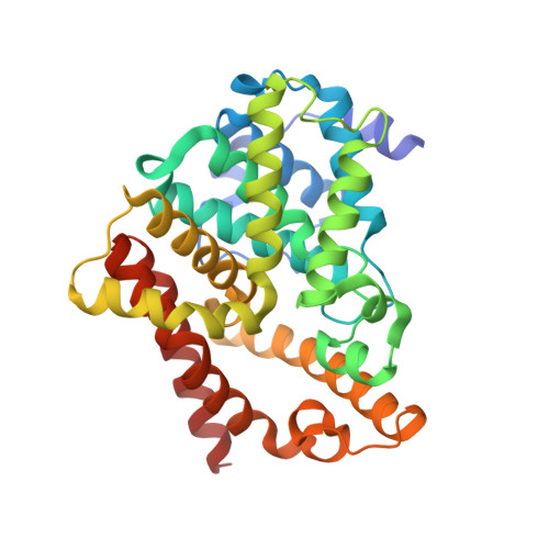
The inhibitory interaction of phosphodiesterase-6 (PDE6) with its gamma-subunit (Pgamma) is pivotal in vertebrate phototransduction. Here, crystal structures of a chimaeric PDE5/PDE6 catalytic domain (PDE5/6cd) complexed with sildenafil or 3-isobutyl-1-methylxanthine and the Pgamma-inhibitory peptide Pgamma(70-87) have been determined at 2.9 and 3.0 A, respectively. These structures show the determinants and the mechanism of the PDE6 inhibition by Pgamma and suggest the conformational change of Pgamma on transducin activation. Two variable H- and M-loops of PDE5/6cd form a distinct interface that contributes to the Pgamma-binding site. This allows the Pgamma C-terminus to fit into the opening of the catalytic pocket, blocking cGMP access to the active site. Our analysis suggests that disruption of the H-M loop interface and Pgamma-binding site is a molecular cause of retinal degeneration in atrd3 mice. Comparison of the two PDE5/6cd structures shows an overlap between the sildenafil and Pgamma(70-87)-binding sites, thereby providing critical insights into the side effects of PDE5 inhibitors on vision.
Organizational Affiliation:
Department of Molecular Physiology and Biophysics, University of Iowa, Iowa City, IA, USA.


