3LEO

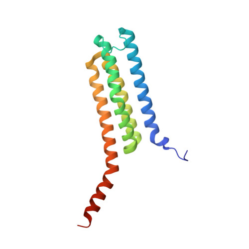
Structure of human Leukotriene C4 synthase mutant R31Q in complex with glutathione


Experimental Data Snapshot

  • Method: X-RAY DIFFRACTION
  • Resolution: 2.10 Å
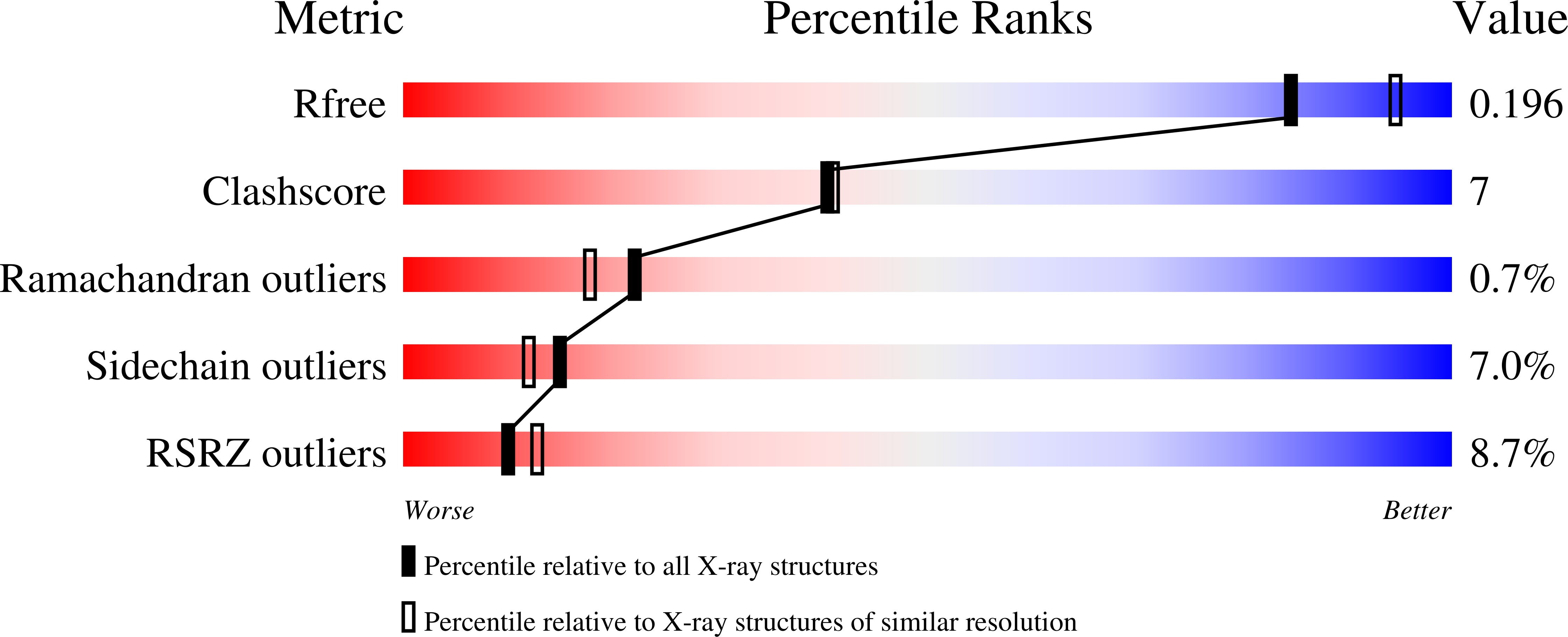
  • R-Value Free: 0.196 
  • R-Value Work: 0.172 
  • R-Value Observed: 0.173 

Starting Model: experimental
View more details

wwPDB Validation   3D Report Full Report


Ligand Structure Quality Assessment 


This is version 1.4 of the entry. See complete history


Literature

Arginine 104 is a key catalytic residue in leukotriene C4 synthase.

Rinaldo-Matthis, A.Wetterholm, A.Martinez Molina, D.Holm, J.Niegowski, D.Ohlson, E.Nordlund, P.Morgenstern, R.Haeggstrom, J.Z.

(2010) J Biol Chem 285: 40771-40776

  • DOI: https://doi.org/10.1074/jbc.M110.105940
  • Primary Citation of Related Structures:  
    3LEO

  • PubMed Abstract: 

    Human leukotriene C(4) synthase (hLTC(4)S) is an integral membrane enzyme that conjugates leukotriene (LT) A(4) with glutathione to form LTC(4), a precursor to the cysteinyl leukotrienes (LTC(4), LTD(4), and LTE(4)) that are involved in the pathogenesis of human bronchial asthma. From the crystal structure of hLTC(4)S, Arg-104 and Arg-31 have been implicated in the conjugation reaction. Here, we used site-directed mutagenesis, UV spectroscopy, and x-ray crystallography to examine the catalytic role of Arg-104 and Arg-31. Exchange of Arg-104 with Ala, Ser, Thr, or Lys abolished 94.3-99.9% of the specific activity against LTA(4). Steady-state kinetics of R104A and R104S revealed that the K(m) for GSH was not significantly affected. UV difference spectra of the binary enzyme-GSH complex indicated that GSH ionization depends on the presence of Arg-104 because no thiolate signal, with λ(max) at 239 nm, could be detected using R104A or R104S hLTC(4)S. Apparently, the interaction of Arg-104 with the thiol group of GSH reduces its pK(a) to allow formation of a thiolate anion and subsequent nucleophilic attack at C6 of LTA(4). On the other hand, exchange of Arg-31 with Ala or Glu reduced the catalytic activity of hLTC(4)S by 88 and 70%, respectively, without significantly affecting the k(cat)/K(m) values for GSH, and a crystal structure of R31Q hLTC(4)S (2.1 Å) revealed a Gln-31 side chain pointing away from the active site. We conclude that Arg-104 plays a critical role in the catalytic mechanism of hLTC(4)S, whereas a functional role of Arg-31 seems more elusive. Because Arg-104 is a conserved residue, our results pertain to other homologous membrane proteins and represent a structure-function paradigm probably common to all microsomal GSH transferases.


  • Organizational Affiliation

    Division of Chemistry 2, Department of Medical Biochemistry and Biophysics, Karolinska Institutet, S-171 77 Stockholm, Sweden.


Macromolecules
Find similar proteins by:  (by identity cutoff)  |  3D Structure
Entity ID: 1
MoleculeChains Sequence LengthOrganismDetailsImage
Leukotriene C4 synthase155Homo sapiensMutation(s): 1 
EC: 4.4.1.20 (PDB Primary Data), 2.5.1 (UniProt)
Membrane Entity: Yes 
UniProt & NIH Common Fund Data Resources
Find proteins for Q16873 (Homo sapiens)
Explore Q16873 
Go to UniProtKB:  Q16873
PHAROS:  Q16873
Entity Groups  
Sequence Clusters30% Identity50% Identity70% Identity90% Identity95% Identity100% Identity
UniProt GroupQ16873
Sequence Annotations
Expand
  • Reference Sequence
Small Molecules
Ligands 7 Unique
IDChains Name / Formula / InChI Key2D Diagram3D Interactions
LMT
Query on LMT

Download Ideal Coordinates CCD File 
N [auth A]DODECYL-BETA-D-MALTOSIDE
C24 H46 O11
NLEBIOOXCVAHBD-QKMCSOCLSA-N
GSH
Query on GSH

Download Ideal Coordinates CCD File 
M [auth A]GLUTATHIONE
C10 H17 N3 O6 S
RWSXRVCMGQZWBV-WDSKDSINSA-N
PLM
Query on PLM

Download Ideal Coordinates CCD File 
C [auth A]
D [auth A]
E [auth A]
G [auth A]
H [auth A]
C [auth A],
D [auth A],
E [auth A],
G [auth A],
H [auth A],
I [auth A]
PALMITIC ACID
C16 H32 O2
IPCSVZSSVZVIGE-UHFFFAOYSA-N
PAM
Query on PAM

Download Ideal Coordinates CCD File 
B [auth A],
P [auth A]
PALMITOLEIC ACID
C16 H30 O2
SECPZKHBENQXJG-FPLPWBNLSA-N
SO4
Query on SO4

Download Ideal Coordinates CCD File 
F [auth A],
J [auth A]
SULFATE ION
O4 S
QAOWNCQODCNURD-UHFFFAOYSA-L
GOL
Query on GOL

Download Ideal Coordinates CCD File 
O [auth A]GLYCEROL
C3 H8 O3
PEDCQBHIVMGVHV-UHFFFAOYSA-N
NI
Query on NI

Download Ideal Coordinates CCD File 
K [auth A],
L [auth A]
NICKEL (II) ION
Ni
VEQPNABPJHWNSG-UHFFFAOYSA-N
Experimental Data & Validation

Experimental Data

  • Method: X-RAY DIFFRACTION
  • Resolution: 2.10 Å
  • R-Value Free: 0.196 
  • R-Value Work: 0.172 
  • R-Value Observed: 0.173 
  • Space Group: F 2 3
Unit Cell:
Length ( Å )Angle ( ˚ )
a = 169.54α = 90
b = 169.54β = 90
c = 169.54γ = 90
Software Package:
Software NamePurpose
XSCALEdata scaling
PHASERphasing
REFMACrefinement
PDB_EXTRACTdata extraction
ADSCdata collection
XDSdata reduction

Structure Validation

View Full Validation Report


Ligand Structure Quality Assessment 


Entry History 

Deposition Data

Revision History  (Full details and data files)

  • Version 1.0: 2010-10-27
    Type: Initial release
  • Version 1.1: 2011-07-13
    Changes: Version format compliance
  • Version 1.2: 2012-03-21
    Changes: Non-polymer description
  • Version 1.3: 2012-12-26
    Changes: Database references
  • Version 1.4: 2023-09-06
    Changes: Data collection, Database references, Derived calculations, Refinement description