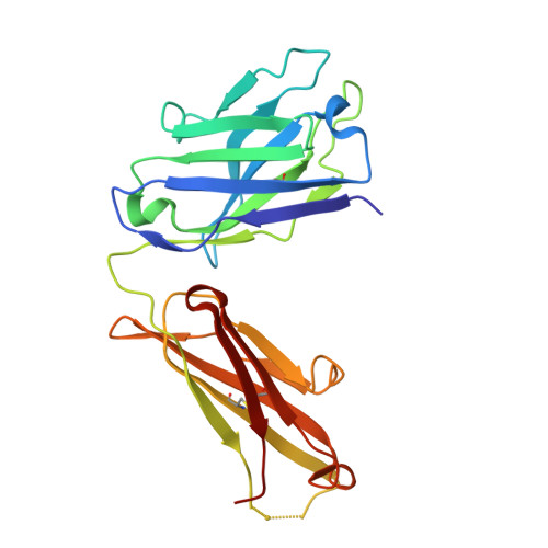
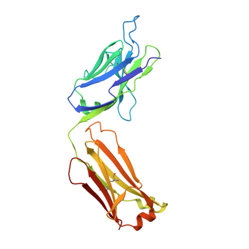
3OKO

Crystal structure of S25-39 in complex with Kdo(2.8)Kdo(2.4)Kdo


Experimental Data Snapshot

  • Method: X-RAY DIFFRACTION
  • Resolution: 2.45 Å
  • R-Value Free: 0.267 
  • R-Value Work: 0.210 
  • R-Value Observed: 0.213 

wwPDB Validation   3D Report Full Report


This is version 2.0 of the entry. See complete history


Literature

A Common NH53K Mutation in the Combining Site of Antibodies Raised against Chlamydial LPS Glycoconjugates Significantly Increases Avidity.

Blackler, R.J.Brooks, C.L.Evans, D.W.Brade, L.Kosma, P.Brade, H.Evans, S.V.

(2011) Biochemistry 50: 3357-3368

  • DOI: https://doi.org/10.1021/bi101886v
  • Primary Citation of Related Structures:  
    3OKD, 3OKE, 3OKK, 3OKL, 3OKM, 3OKN, 3OKO

  • PubMed Abstract: 

    The crystal structures of the antigen-binding fragment of the murine monoclonal antibody (mAb) S25-39 in the presence of several antigens representing chlamydial lipopolysaccharide (LPS) epitopes based on the bacterial sugar 3-deoxy-α-D-manno-oct-2-ulosonic acid (Kdo) have been determined at resolutions from 2.4 to 1.8 Å. The antigen-binding site of this antibody differs from the well-characterized antibody S25-2 by a single mutation away from the germline of asparagine H53 to lysine, yet this one mutation results in a significant increase in avidity across a range of antigens. A comparison of the two antibody structures reveals that the mutated Lys H53 forms additional hydrogen bonds and/or charged-residue interactions with the second Kdo residue of every antigen having two or more carbohydrate residues. Significantly, the NH53K mutation results from a single nucleotide substitution in the germline sequence common among a panel of antibodies raised against glycoconjugates containing carbohydrate epitopes of chlamydial LPS. Like S25-2, S25-39 displays significant induced fit of complementarity determining region (CDR) H3 upon antigen binding, with the unliganded structure possessing a conformation distinct from those reported earlier for S25-2. The four different observed conformations for CDR H3 suggest that this CDR has evolved to exploit the recognition potential of a flexible loop while minimizing the associated entropic penalties of binding by adopting a limited number of ordered conformations in the unliganded state. These observations reveal strategies evolved to balance adaptability and specificity in the germline antibody response to carbohydrate antigens.


  • Organizational Affiliation

    Department of Biochemistry and Microbiology, University of Victoria , Victoria, BC V8P 3P6, Canada.


Macromolecules
Find similar proteins by:  (by identity cutoff)  |  3D Structure
Entity ID: 1
MoleculeChains Sequence LengthOrganismDetailsImage
S25-39 Fab (IgG1k) light chainA [auth B]222Mus musculusMutation(s): 0 
Entity Groups  
Sequence Clusters30% Identity50% Identity70% Identity90% Identity95% Identity100% Identity
Sequence Annotations
Expand
  • Reference Sequence
Find similar proteins by:  (by identity cutoff)  |  3D Structure
Entity ID: 2
MoleculeChains Sequence LengthOrganismDetailsImage
S25-39 Fab (IgG1k) heavy chainB [auth A]219Mus musculusMutation(s): 0 
Entity Groups  
Sequence Clusters30% Identity50% Identity70% Identity90% Identity95% Identity100% Identity
Sequence Annotations
Expand
  • Reference Sequence
Oligosaccharides

Help

Entity ID: 3
MoleculeChains Length2D Diagram Glycosylation3D Interactions
3-deoxy-alpha-D-manno-oct-2-ulopyranosonic acid-(2-8)-3-deoxy-alpha-D-manno-oct-2-ulopyranosonic acid-(2-4)-prop-2-en-1-yl 3-deoxy-alpha-D-manno-oct-2-ulopyranosidonic acid
C
3N/AN/A
Glycosylation Resources
GlyTouCan:  G25386BJ
GlyCosmos:  G25386BJ
Small Molecules
Ligands 1 Unique
IDChains Name / Formula / InChI Key2D Diagram3D Interactions
ZN
Query on ZN

Download Ideal Coordinates CCD File 
D [auth A]ZINC ION
Zn
PTFCDOFLOPIGGS-UHFFFAOYSA-N
Experimental Data & Validation

Experimental Data

  • Method: X-RAY DIFFRACTION
  • Resolution: 2.45 Å
  • R-Value Free: 0.267 
  • R-Value Work: 0.210 
  • R-Value Observed: 0.213 
  • Space Group: P 21 21 21
Unit Cell:
Length ( Å )Angle ( ˚ )
a = 45.441α = 90
b = 81.394β = 90
c = 129.009γ = 90
Software Package:
Software NamePurpose
d*TREKdata scaling
PHASERphasing
PHENIXrefinement
PDB_EXTRACTdata extraction

Structure Validation

View Full Validation Report


Entry History 

Deposition Data

Revision History  (Full details and data files)

  • Version 1.0: 2011-04-06
    Type: Initial release
  • Version 1.1: 2011-07-13
    Changes: Version format compliance
  • Version 1.2: 2018-03-07
    Changes: Data collection
  • Version 2.0: 2020-07-29
    Type: Remediation
    Reason: Carbohydrate remediation
    Changes: Atomic model, Data collection, Database references, Derived calculations, Structure summary