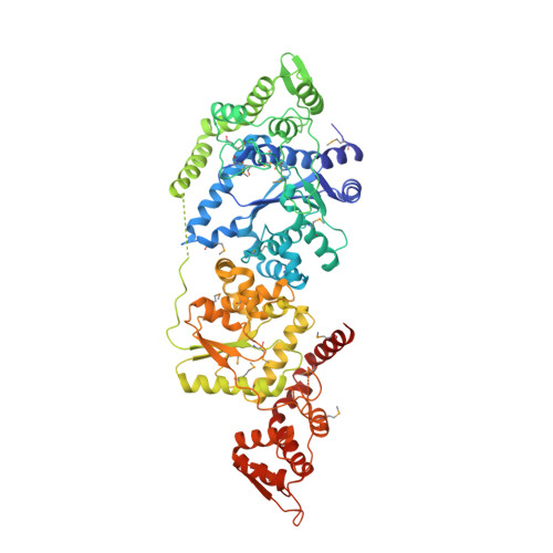
3P0H

Leishmania major Tyrosyl-tRNA synthetase in complex with fisetin, cubic crystal form


Experimental Data Snapshot

  • Method: X-RAY DIFFRACTION
  • Resolution: 3.00 Å
  • R-Value Free: 0.273 
  • R-Value Work: 0.234 
  • R-Value Observed: 0.235 

Starting Models: experimental
View more details

wwPDB Validation   3D Report Full Report


Ligand Structure Quality Assessment 


This is version 1.4 of the entry. See complete history


Literature

The Double-Length Tyrosyl-tRNA Synthetase from the Eukaryote Leishmania major Forms an Intrinsically Asymmetric Pseudo-Dimer.

Larson, E.T.Kim, J.E.Castaneda, L.J.Napuli, A.J.Zhang, Z.Fan, E.Zucker, F.H.Verlinde, C.L.Buckner, F.S.Van Voorhis, W.C.Hol, W.G.Merritt, E.A.

(2011) J Mol Biol 409: 159-176

  • DOI: https://doi.org/10.1016/j.jmb.2011.03.026
  • Primary Citation of Related Structures:  
    3P0H, 3P0I, 3P0J

  • PubMed Abstract: 

    The single tyrosyl-tRNA synthetase (TyrRS) gene in trypanosomatid genomes codes for a protein that is twice the length of TyrRS from virtually all other organisms. Each half of the double-length TyrRS contains a catalytic domain and an anticodon-binding domain; however, the two halves retain only 17% sequence identity to each other. The structural and functional consequences of this duplication and divergence are unclear. TyrRS normally forms a homodimer in which the active site of one monomer pairs with the anticodon-binding domain from the other. However, crystal structures of Leishmania major TyrRS show that, instead, the two halves of a single molecule form a pseudo-dimer resembling the canonical TyrRS dimer. Curiously, the C-terminal copy of the catalytic domain has lost the catalytically important HIGH and KMSKS motifs characteristic of class I aminoacyl-tRNA synthetases. Thus, the pseudo-dimer contains only one functional active site (contributed by the N-terminal half) and only one functional anticodon recognition site (contributed by the C-terminal half). Despite biochemical evidence for negative cooperativity between the two active sites of the usual TyrRS homodimer, previous structures have captured a crystallographically-imposed symmetric state. As the L. major TyrRS pseudo-dimer is inherently asymmetric, conformational variations observed near the active site may be relevant to understanding how the state of a single active site is communicated across the dimer interface. Furthermore, substantial differences between trypanosomal TyrRS and human homologs are promising for the design of inhibitors that selectively target the parasite enzyme.


  • Organizational Affiliation

    Medical Structural Genomics of Pathogenic Protozoa, Department of Biochemistry, University of Washington, Seattle, WA 98195, USA.


Macromolecules
Find similar proteins by:  (by identity cutoff)  |  3D Structure
Entity ID: 1
MoleculeChains Sequence LengthOrganismDetailsImage
Tyrosyl-tRNA synthetase
A, B
690Leishmania majorMutation(s): 0 
Gene Names: LmjF14.1370
EC: 6.1.1.1
UniProt
Find proteins for Q4QFJ7 (Leishmania major)
Explore Q4QFJ7 
Go to UniProtKB:  Q4QFJ7
Entity Groups  
Sequence Clusters30% Identity50% Identity70% Identity90% Identity95% Identity100% Identity
UniProt GroupQ4QFJ7
Sequence Annotations
Expand
  • Reference Sequence
Small Molecules
Ligands 1 Unique
IDChains Name / Formula / InChI Key2D Diagram3D Interactions
FSE
Query on FSE

Download Ideal Coordinates CCD File 
C [auth A],
D [auth B]
3,7,3',4'-TETRAHYDROXYFLAVONE
C15 H10 O6
XHEFDIBZLJXQHF-UHFFFAOYSA-N
Modified Residues  1 Unique
IDChains TypeFormula2D DiagramParent
MSE
Query on MSE
A, B
L-PEPTIDE LINKINGC5 H11 N O2 SeMET
Experimental Data & Validation

Experimental Data

  • Method: X-RAY DIFFRACTION
  • Resolution: 3.00 Å
  • R-Value Free: 0.273 
  • R-Value Work: 0.234 
  • R-Value Observed: 0.235 
  • Space Group: I 2 3
Unit Cell:
Length ( Å )Angle ( ˚ )
a = 240.24α = 90
b = 240.24β = 90
c = 240.24γ = 90
Software Package:
Software NamePurpose
MOSFLMdata reduction
SCALAdata scaling
RESOLVEphasing
REFMACrefinement
PDB_EXTRACTdata extraction
Blu-Icedata collection
BALBESphasing
PHASERphasing

Structure Validation

View Full Validation Report


Ligand Structure Quality Assessment 


Entry History 

Deposition Data

Revision History  (Full details and data files)

  • Version 1.0: 2011-03-23
    Type: Initial release
  • Version 1.1: 2011-07-13
    Changes: Version format compliance
  • Version 1.2: 2017-11-08
    Changes: Refinement description
  • Version 1.3: 2023-09-06
    Changes: Data collection, Database references, Derived calculations, Refinement description, Structure summary
  • Version 1.4: 2023-12-06
    Changes: Data collection