3RUB

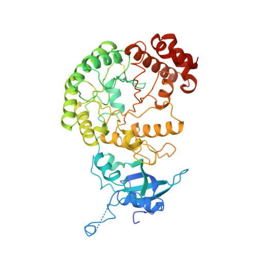
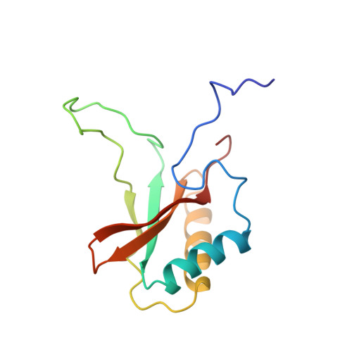
CRYSTAL STRUCTURE OF THE UNACTIVATED FORM OF RIBULOSE-1,5-BISPHOSPHATE CARBOXYLASE(SLASH)OXYGENASE FROM TOBACCO REFINED AT 2.0-ANGSTROMS RESOLUTION


Experimental Data Snapshot

  • Method: X-RAY DIFFRACTION
  • Resolution: 2.00 Å
  • R-Value Observed: 0.171 

wwPDB Validation   3D Report Full Report


This is version 1.4 of the entry. See complete history


Literature

Crystal structure of the unactivated form of ribulose-1,5-bisphosphate carboxylase/oxygenase from tobacco refined at 2.0-A resolution.

Curmi, P.M.Cascio, D.Sweet, R.M.Eisenberg, D.Schreuder, H.

(1992) J Biol Chem 267: 16980-16989

  • Primary Citation of Related Structures:  
    3RUB

  • PubMed Abstract: 

    The structure of the unactivated form of ribulose-1,5-bisphosphate carboxylase/oxygenase was refined at a resolution of 2.0 A to an R-factor of 17.1%. The previous model (Chapman et al., 1988) was extensively rebuilt, and the small subunit was retraced. The refined model consists of residues 22-63 and 69-467 of the large subunit and the complete small subunit. A striking feature of the model is that several loops have very high B-factors, probably representing mobile regions of the molecule. An examination of the intersubunit contacts shows that the L8S8 hexadecamer is composed of four L2 dimers. The dominant contacts between these L2 dimers are formed by the small subunits. This suggests that the small subunits may be essential for maintaining the integrity of the L8S8 structure. The active site shows differences between the unactivated form and the quaternary complex. In particular, Lys334 has moved out of the active site by about 10A. This residue lies on loop 6 of the alpha beta barrel, which is a particularly mobile loop. The site of ribulose-1,5-bisphosphate carboxylase/oxygenase activation is well ordered in the absence of the carbamylation of Lys201 and Mg2+ binding. The residues are held poised by a network of hydrogen bonds. In the unactivated state, the active site is accessible to substrate binding.


  • Organizational Affiliation

    Department of Chemistry and Biochemistry, University of California, Los Angeles 90024.


Macromolecules
Find similar proteins by:  (by identity cutoff)  |  3D Structure
Entity ID: 1
MoleculeChains Sequence LengthOrganismDetailsImage
RIBULOSE 1,5-BISPHOSPHATE CARBOXYLASE/OXYGENASE, FORM IIIA [auth L]477Nicotiana tabacumMutation(s): 0 
EC: 4.1.1.39
UniProt
Find proteins for P00876 (Nicotiana tabacum)
Explore P00876 
Go to UniProtKB:  P00876
Entity Groups  
Sequence Clusters30% Identity50% Identity70% Identity90% Identity95% Identity100% Identity
UniProt GroupP00876
Sequence Annotations
Expand
  • Reference Sequence
Find similar proteins by:  (by identity cutoff)  |  3D Structure
Entity ID: 2
MoleculeChains Sequence LengthOrganismDetailsImage
RIBULOSE 1,5-BISPHOSPHATE CARBOXYLASE/OXYGENASE, FORM IIIB [auth S]123Nicotiana tabacumMutation(s): 0 
Gene Names: RBCS
EC: 4.1.1.39
UniProt
Find proteins for P69249 (Nicotiana tabacum)
Explore P69249 
Go to UniProtKB:  P69249
Entity Groups  
Sequence Clusters30% Identity50% Identity70% Identity90% Identity95% Identity100% Identity
UniProt GroupP69249
Sequence Annotations
Expand
  • Reference Sequence
Experimental Data & Validation

Experimental Data

  • Method: X-RAY DIFFRACTION
  • Resolution: 2.00 Å
  • R-Value Observed: 0.171 
  • Space Group: I 4 2 2
Unit Cell:
Length ( Å )Angle ( ˚ )
a = 148.7α = 90
b = 148.7β = 90
c = 137.5γ = 90
Software Package:
Software NamePurpose
PROFFTrefinement

Structure Validation

View Full Validation Report


Entry History 

Deposition Data

Revision History  (Full details and data files)

  • Version 1.0: 1992-10-15
    Type: Initial release
  • Version 1.1: 2008-03-03
    Changes: Version format compliance
  • Version 1.2: 2011-07-13
    Changes: Derived calculations, Version format compliance
  • Version 1.3: 2024-06-05
    Changes: Data collection, Database references, Derived calculations, Other
  • Version 1.4: 2024-10-23
    Changes: Structure summary