3ZCH

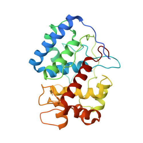
Ascorbate peroxidase W41A-H42M mutant


Experimental Data Snapshot

  • Method: X-RAY DIFFRACTION
  • Resolution: 2.00 Å
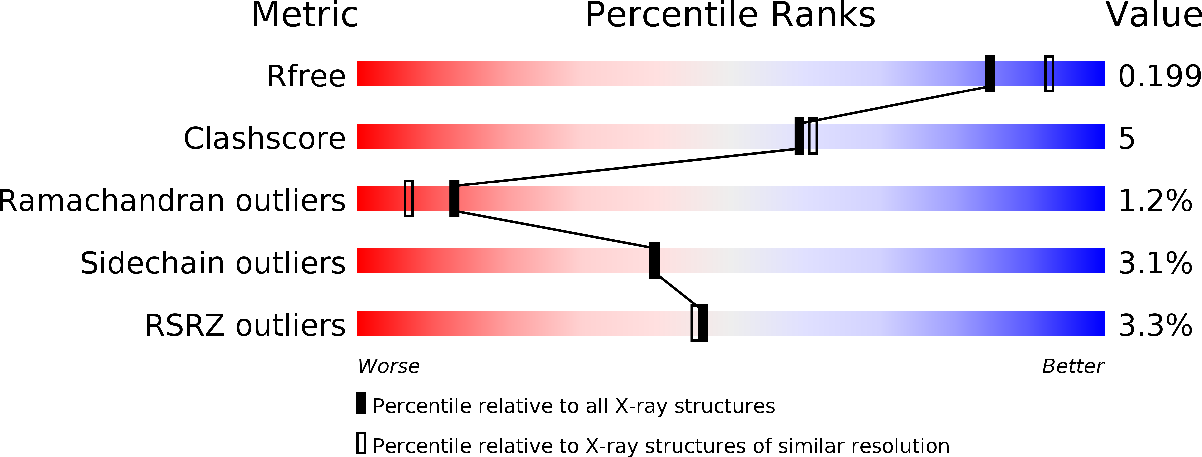
  • R-Value Free: 0.199 
  • R-Value Work: 0.153 
  • R-Value Observed: 0.155 

Starting Model: experimental
View more details

wwPDB Validation   3D Report Full Report


Ligand Structure Quality Assessment 


This is version 1.2 of the entry. See complete history


Literature

Probing the Conformational Mobility of the Active Site of Ascorbate Peroxidase

Guimero, A.Badyal, S.K.Leeks, T.Moody, P.C.E.Raven, E.L.

(2013) Dalton Trans 42: 3170

  • DOI: https://doi.org/10.1039/c2dt32455e
  • Primary Citation of Related Structures:  
    3ZCG, 3ZCH, 3ZCY

  • PubMed Abstract: 

    We have previously demonstrated (Badyal et al., J. Biol. Chem., 2006, 281, 24512) that removal of the active site tryptophan (Trp41) in ascorbate peroxidase increases the conformational mobility of the distal histidine residue (His42) and that His42 coordinates to the iron in the oxidised W41A enzyme to give a 6-coordinate, low-spin peroxidase. In this work, we probe the conformational flexibility of the active site in more detail. We examine whether other residues (Cys, Tyr, Met) can also ligate to the heme at position 42; we find that introduction of other ligating amino acids created a cavity in the heme pocket, but that formation of 6-coordinate heme is not observed. In addition, we examine the role of Asn-71, which hydrogen bonds to His42 and tethers the distal histidine in the active site pocket; we find that removal of this hydrogen bond increases the proportion of low-spin heme. We suggest that, in addition to its well-known role in facilitating the reaction with peroxide, His42 also plays a role in defining the shape and folding of the active site pocket.


Macromolecules
Find similar proteins by:  (by identity cutoff)  |  3D Structure
Entity ID: 1
MoleculeChains Sequence LengthOrganismDetailsImage
ASCORBATE PEROXIDASE261Glycine maxMutation(s): 2 
EC: 1.11.1.11
UniProt
Find proteins for Q43758 (Glycine max)
Explore Q43758 
Go to UniProtKB:  Q43758
Entity Groups  
Sequence Clusters30% Identity50% Identity70% Identity90% Identity95% Identity100% Identity
UniProt GroupQ43758
Sequence Annotations
Expand
  • Reference Sequence
Experimental Data & Validation

Experimental Data

  • Method: X-RAY DIFFRACTION
  • Resolution: 2.00 Å
  • R-Value Free: 0.199 
  • R-Value Work: 0.153 
  • R-Value Observed: 0.155 
  • Space Group: P 42 21 2
Unit Cell:
Length ( Å )Angle ( ˚ )
a = 82.629α = 90
b = 82.629β = 90
c = 74.95γ = 90
Software Package:
Software NamePurpose
PHENIXrefinement
XDSdata reduction
SCALAdata scaling
MOLREPphasing

Structure Validation

View Full Validation Report


Ligand Structure Quality Assessment 


Entry History 

Deposition Data

Revision History  (Full details and data files)

  • Version 1.0: 2012-12-19
    Type: Initial release
  • Version 1.1: 2013-02-27
    Changes: Database references
  • Version 1.2: 2023-12-20
    Changes: Data collection, Database references, Derived calculations, Other, Refinement description