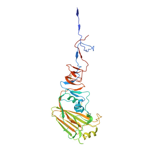
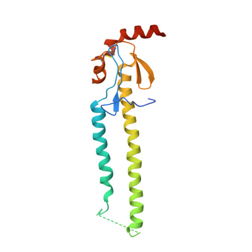
4EDA

Structures of monomeric hemagglutinin and its complex with an Fab fragment of a neutralizing antibody that binds to H1 subtype influenza viruses: molecular basis of infectivity of 2009 pandemic H1N1 influenza A viruses


Experimental Data Snapshot

  • Method: X-RAY DIFFRACTION
  • Resolution: 2.70 Å
  • R-Value Free: 0.289 
  • R-Value Work: 0.237 
  • R-Value Observed: 0.240 

wwPDB Validation   3D Report Full Report


Ligand Structure Quality Assessment 


This is version 2.0 of the entry. See complete history


Literature

Insight into structural diversity of influenza virus haemagglutinin

Cho, K.J.Lee, J.H.Hong, K.W.Kim, S.H.Park, Y.Lee, J.Y.Kang, S.Kim, S.Yang, J.H.Kim, E.K.Seok, J.H.Unzai, S.Park, S.Y.Saelens, X.Kim, C.J.Lee, J.Y.Kang, C.Oh, H.B.Chung, M.S.Kim, K.H.

(2013) J Gen Virol 94: 1712-1722

  • DOI: https://doi.org/10.1099/vir.0.051136-0
  • Primary Citation of Related Structures:  
    4EDA, 4EDB

  • PubMed Abstract: 

    Influenza virus infects host cells through membrane fusion, a process mediated by the low pH-induced conformational change of the viral surface glycoprotein haemagglutinin (HA). We determined the structures and biochemical properties of the HA proteins from A/Korea/01/2009 (KR01), a 2009 pandemic strain, and A/Thailand/CU44/2006 (CU44), a seasonal strain. The crystal structure of KR01 HA revealed a V-shaped head-to-head arrangement, which is not seen in other HA proteins including CU44 HA. We isolated a broadly neutralizing H1-specific monoclonal antibody GC0757. The KR01 HA-Fab0757 complex structure also exhibited a head-to-head arrangement of HA. Both native and Fab complex structures reveal a different spatial orientation of HA1 relative to HA2, indicating that HA is flexible and dynamic at neutral pH. Further, the KR01 HA exhibited significantly lower protein stability and increased susceptibility to proteolytic cleavage compared with other HAs. Our structures provide important insights into the conformational flexibility of HA.


  • Organizational Affiliation

    Department of Biotechnology & Bioinformatics, College of Science & Technology, Korea University, Sejong 339-700, Korea.


Macromolecules
Find similar proteins by:  (by identity cutoff)  |  3D Structure
Entity ID: 1
MoleculeChains Sequence LengthOrganismDetailsImage
Hemagglutinin
A, C
336Influenza A virus (A/Korea/01/2009(H1N1))Mutation(s): 0 
Gene Names: HA
UniProt
Find proteins for C5MQE6 (Influenza A virus)
Explore C5MQE6 
Go to UniProtKB:  C5MQE6
Entity Groups  
Sequence Clusters30% Identity50% Identity70% Identity90% Identity95% Identity100% Identity
UniProt GroupC5MQE6
Glycosylation
Glycosylation Sites: 3
Sequence Annotations
Expand
  • Reference Sequence
Find similar proteins by:  (by identity cutoff)  |  3D Structure
Entity ID: 2
MoleculeChains Sequence LengthOrganismDetailsImage
Hemagglutinin
B, D
182Influenza A virus (A/Korea/01/2009(H1N1))Mutation(s): 0 
Gene Names: HA
UniProt
Find proteins for C5MQE6 (Influenza A virus)
Explore C5MQE6 
Go to UniProtKB:  C5MQE6
Entity Groups  
Sequence Clusters30% Identity50% Identity70% Identity90% Identity95% Identity100% Identity
UniProt GroupC5MQE6
Sequence Annotations
Expand
  • Reference Sequence
Oligosaccharides

Help

Entity ID: 3
MoleculeChains Length2D Diagram Glycosylation3D Interactions
2-acetamido-2-deoxy-beta-D-glucopyranose-(1-4)-2-acetamido-2-deoxy-beta-D-glucopyranose
E, F
2N-Glycosylation
Glycosylation Resources
GlyTouCan:  G42666HT
GlyCosmos:  G42666HT
GlyGen:  G42666HT
Experimental Data & Validation

Experimental Data

  • Method: X-RAY DIFFRACTION
  • Resolution: 2.70 Å
  • R-Value Free: 0.289 
  • R-Value Work: 0.237 
  • R-Value Observed: 0.240 
  • Space Group: P 6
Unit Cell:
Length ( Å )Angle ( ˚ )
a = 208.126α = 90
b = 208.126β = 90
c = 65.765γ = 120
Software Package:
Software NamePurpose
DENZOdata reduction
SCALEPACKdata scaling
PHENIXrefinement
PDB_EXTRACTdata extraction
HKL-2000data collection
PHASERphasing

Structure Validation

View Full Validation Report


Ligand Structure Quality Assessment 


Entry History 

Deposition Data

Revision History  (Full details and data files)

  • Version 1.0: 2013-05-22
    Type: Initial release
  • Version 1.1: 2014-03-12
    Changes: Database references, Derived calculations
  • Version 1.2: 2017-11-15
    Changes: Refinement description
  • Version 2.0: 2020-07-29
    Type: Remediation
    Reason: Carbohydrate remediation
    Changes: Advisory, Atomic model, Data collection, Database references, Derived calculations, Structure summary