4FAJ

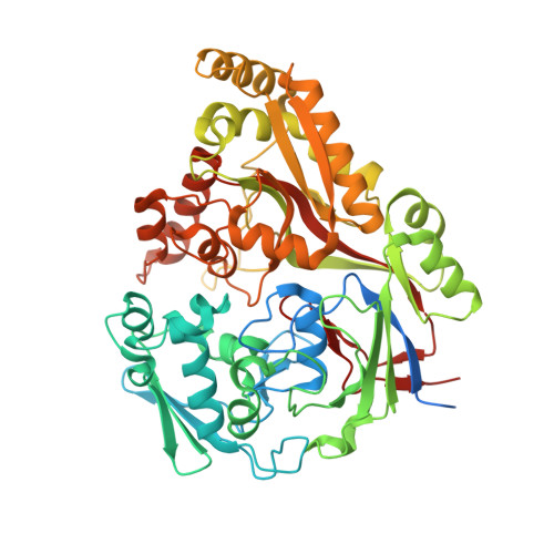
Structure and mode of peptide binding of pheromone receptor PrgZ


Experimental Data Snapshot

  • Method: X-RAY DIFFRACTION
  • Resolution: 1.90 Å
  • R-Value Free: 0.193 
  • R-Value Work: 0.156 
  • R-Value Observed: 0.158 

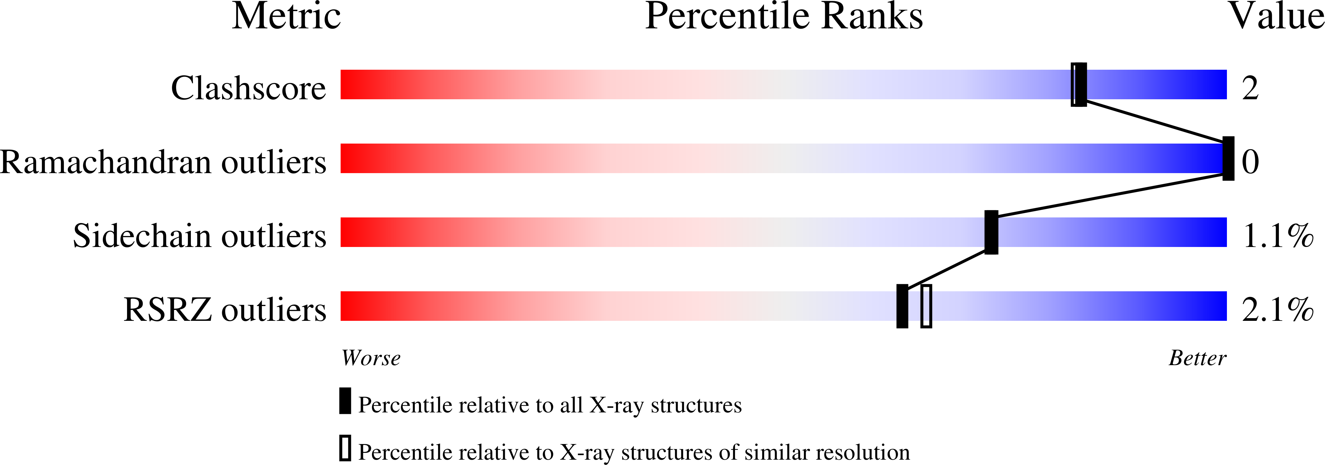
wwPDB Validation   3D Report Full Report


This is version 1.4 of the entry. See complete history


Literature

Structure and Mode of Peptide Binding of Pheromone Receptor PrgZ.

Berntsson, R.P.Schuurman-Wolters, G.K.Dunny, G.Slotboom, D.J.Poolman, B.

(2012) J Biol Chem 287: 37165-37170

  • DOI: https://doi.org/10.1074/jbc.M112.386334
  • Primary Citation of Related Structures:  
    4FAJ

  • PubMed Abstract: 

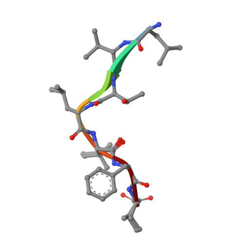
    We present the crystal structure of the pheromone receptor protein PrgZ from Enterococcus faecalis in complex with the heptapeptide cCF10 (LVTLVFV), which is used in signaling between conjugative recipient and donor cells. Comparison of PrgZ with homologous oligopeptide-binding proteins (AppA and OppA) explains the high specificity of PrgZ for hydrophobic heptapeptides versus the promiscuity of peptide binding in the homologous proteins.


  • Organizational Affiliation

    Department of Biochemistry, Groningen Biomolecular Sciences and Biotechnology Institute, Netherlands Proteomics Centre, University of Groningen, 9747 AG Groningen, The Netherlands.


Macromolecules
Find similar proteins by:  (by identity cutoff)  |  3D Structure
Entity ID: 1
MoleculeChains Sequence LengthOrganismDetailsImage
PrgZ564Enterococcus faecalisMutation(s): 0 
Gene Names: prgZ
UniProt
Find proteins for Q51643 (Enterococcus faecalis)
Explore Q51643 
Go to UniProtKB:  Q51643
Entity Groups  
Sequence Clusters30% Identity50% Identity70% Identity90% Identity95% Identity100% Identity
UniProt GroupQ51643
Sequence Annotations
Expand
  • Reference Sequence

Find similar proteins by:  Sequence   |   3D Structure  

Entity ID: 2
MoleculeChains Sequence LengthOrganismDetailsImage
Sex pheromone cCF107Enterococcus faecalisMutation(s): 0 
UniProt
Find proteins for P20104 (Enterococcus faecalis)
Explore P20104 
Go to UniProtKB:  P20104
Entity Groups  
UniProt GroupP20104
Sequence Annotations
Expand
  • Reference Sequence
Small Molecules
Ligands 2 Unique
IDChains Name / Formula / InChI Key2D Diagram3D Interactions
GOL
Query on GOL

Download Ideal Coordinates CCD File 
C [auth A],
D [auth A],
I [auth A],
J [auth A]
GLYCEROL
C3 H8 O3
PEDCQBHIVMGVHV-UHFFFAOYSA-N
ZN
Query on ZN

Download Ideal Coordinates CCD File 
E [auth A],
F [auth A],
G [auth A],
H [auth A]
ZINC ION
Zn
PTFCDOFLOPIGGS-UHFFFAOYSA-N
Experimental Data & Validation

Experimental Data

  • Method: X-RAY DIFFRACTION
  • Resolution: 1.90 Å
  • R-Value Free: 0.193 
  • R-Value Work: 0.156 
  • R-Value Observed: 0.158 
  • Space Group: P 21 21 21
Unit Cell:
Length ( Å )Angle ( ˚ )
a = 68.622α = 90
b = 74.122β = 90
c = 112.622γ = 90
Software Package:
Software NamePurpose
XSCALEdata scaling
PHASERphasing
REFMACrefinement
PDB_EXTRACTdata extraction

Structure Validation

View Full Validation Report


Entry History 

Deposition Data

Revision History  (Full details and data files)

  • Version 1.0: 2012-09-12
    Type: Initial release
  • Version 1.1: 2012-11-14
    Changes: Database references, Derived calculations
  • Version 1.2: 2012-12-12
    Changes: Derived calculations
  • Version 1.3: 2018-01-24
    Changes: Structure summary
  • Version 1.4: 2024-02-28
    Changes: Data collection, Database references, Derived calculations