4FHE

Spore photoproduct lyase C140A mutant


Experimental Data Snapshot

  • Method: X-RAY DIFFRACTION
  • Resolution: 2.00 Å
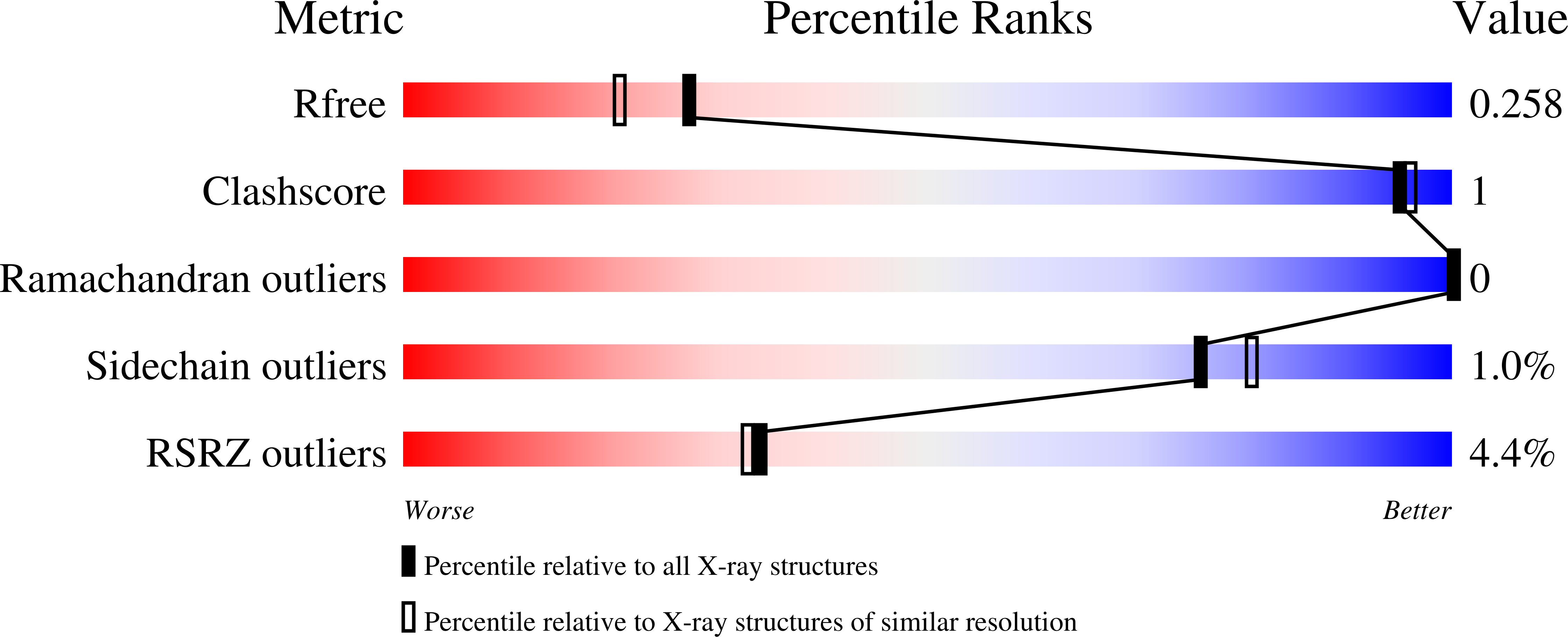
  • R-Value Free: 0.256 
  • R-Value Work: 0.221 
  • R-Value Observed: 0.223 

Starting Model: experimental
View more details

wwPDB Validation   3D Report Full Report


Ligand Structure Quality Assessment 


This is version 1.3 of the entry. See complete history


Literature

Structural insights into recognition and repair of UV-DNA damage by Spore Photoproduct Lyase, a radical SAM enzyme.

Benjdia, A.Heil, K.Barends, T.R.Carell, T.Schlichting, I.

(2012) Nucleic Acids Res 40: 9308-9318

  • DOI: https://doi.org/10.1093/nar/gks603
  • Primary Citation of Related Structures:  
    4FHC, 4FHD, 4FHE, 4FHF, 4FHG

  • PubMed Abstract: 

    Bacterial spores possess an enormous resistance to ultraviolet (UV) radiation. This is largely due to a unique DNA repair enzyme, Spore Photoproduct Lyase (SP lyase) that repairs a specific UV-induced DNA lesion, the spore photoproduct (SP), through an unprecedented radical-based mechanism. Unlike DNA photolyases, SP lyase belongs to the emerging superfamily of radical S-adenosyl-l-methionine (SAM) enzymes and uses a [4Fe-4S](1+) cluster and SAM to initiate the repair reaction. We report here the first crystal structure of this enigmatic enzyme in complex with its [4Fe-4S] cluster and its SAM cofactor, in the absence and presence of a DNA lesion, the dinucleoside SP. The high resolution structures provide fundamental insights into the active site, the DNA lesion recognition and binding which involve a β-hairpin structure. We show that SAM and a conserved cysteine residue are perfectly positioned in the active site for hydrogen atom abstraction from the dihydrothymine residue of the lesion and donation to the α-thyminyl radical moiety, respectively. Based on structural and biochemical characterizations of mutant proteins, we substantiate the role of this cysteine in the enzymatic mechanism. Our structure reveals how SP lyase combines specific features of radical SAM and DNA repair enzymes to enable a complex radical-based repair reaction to take place.


  • Organizational Affiliation

    Department of Biomolecular Mechanisms, Max-Planck Institute for Medical Research, Jahnstrasse 29, 69120 Heidelberg, Germany. Alhosna.Benjdia@mpimf-heidelberg.mpg.de


Macromolecules
Find similar proteins by:  (by identity cutoff)  |  3D Structure
Entity ID: 1
MoleculeChains Sequence LengthOrganismDetailsImage
Spore photoproduct lyase368Geobacillus thermodenitrificans NG80-2Mutation(s): 1 
Gene Names: GTNG_2348
UniProt
Find proteins for A4IQU1 (Geobacillus thermodenitrificans (strain NG80-2))
Explore A4IQU1 
Go to UniProtKB:  A4IQU1
Entity Groups  
Sequence Clusters30% Identity50% Identity70% Identity90% Identity95% Identity100% Identity
UniProt GroupA4IQU1
Sequence Annotations
Expand
  • Reference Sequence
Small Molecules
Ligands 4 Unique
IDChains Name / Formula / InChI Key2D Diagram3D Interactions
EEM
Query on EEM

Download Ideal Coordinates CCD File 
H [auth A][(3S)-3-amino-4-hydroxy-4-oxo-butyl]-[[(2S,3S,4R,5R)-5-(6-aminopurin-9-yl)-3,4-dihydroxy-oxolan-2-yl]methyl]-methyl-selanium
C15 H23 N6 O5 Se
GGJFWMOVUFBSIN-FCKMPRQPSA-O
SF4
Query on SF4

Download Ideal Coordinates CCD File 
B [auth A]IRON/SULFUR CLUSTER
Fe4 S4
LJBDFODJNLIPKO-UHFFFAOYSA-N
SO4
Query on SO4

Download Ideal Coordinates CCD File 
C [auth A],
D [auth A],
E [auth A],
F [auth A]
SULFATE ION
O4 S
QAOWNCQODCNURD-UHFFFAOYSA-L
EDO
Query on EDO

Download Ideal Coordinates CCD File 
G [auth A]1,2-ETHANEDIOL
C2 H6 O2
LYCAIKOWRPUZTN-UHFFFAOYSA-N
Experimental Data & Validation

Experimental Data

  • Method: X-RAY DIFFRACTION
  • Resolution: 2.00 Å
  • R-Value Free: 0.256 
  • R-Value Work: 0.221 
  • R-Value Observed: 0.223 
  • Space Group: P 21 21 21
Unit Cell:
Length ( Å )Angle ( ˚ )
a = 54.02α = 90
b = 63.03β = 90
c = 143.62γ = 90
Software Package:
Software NamePurpose
HEIDIdata collection
PHASERphasing
REFMACrefinement
XDSdata reduction
XSCALEdata scaling

Structure Validation

View Full Validation Report


Ligand Structure Quality Assessment 


Entry History 

Deposition Data

Revision History  (Full details and data files)

  • Version 1.0: 2012-07-18
    Type: Initial release
  • Version 1.1: 2012-10-31
    Changes: Database references
  • Version 1.2: 2024-02-28
    Changes: Data collection, Database references, Derived calculations
  • Version 1.3: 2024-04-03
    Changes: Refinement description