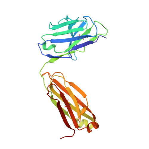
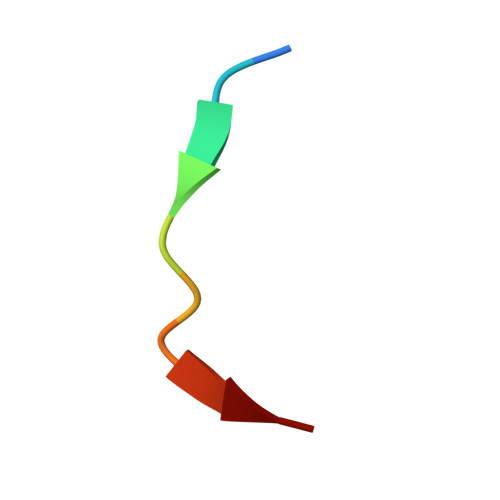
4HHA

Anti-Human Cytomegalovirus (HCMV) Fab KE5 with epitope peptide AD-2S1


Experimental Data Snapshot

  • Method: X-RAY DIFFRACTION
  • Resolution: 1.60 Å
  • R-Value Free: 0.226 
  • R-Value Work: 0.213 
  • R-Value Observed: 0.213 

Starting Model: experimental
View more details

wwPDB Validation   3D Report Full Report


This is version 1.3 of the entry. See complete history


Literature

Structures of Preferred Human IgV Genes-Based Protective Antibodies Identify How Conserved Residues Contact Diverse Antigens and Assign Source of Specificity to CDR3 Loop Variation.

Bryson, S.Thomson, C.A.Risnes, L.F.Dasgupta, S.Smith, K.Schrader, J.W.Pai, E.F.

(2016) J Immunol 196: 4723-4730

  • DOI: https://doi.org/10.4049/jimmunol.1402890
  • Primary Citation of Related Structures:  
    4HH9, 4HHA, 4HIE, 4HIH, 4HII, 4HIJ, 4PTT, 4PTU

  • PubMed Abstract: 

    The human Ab response to certain pathogens is oligoclonal, with preferred IgV genes being used more frequently than others. A pair of such preferred genes, IGVK3-11 and IGVH3-30, contributes to the generation of protective Abs directed against the 23F serotype of the pneumonococcal capsular polysaccharide of Streptococcus pneumoniae and against the AD-2S1 peptide of the gB membrane protein of human CMV. Structural analyses of Fab fragments of mAbs 023.102 and pn132p2C05 in complex with portions of the 23F polysaccharide revealed five germline-encoded residues in contact with the key component, l-rhamnose. In the case of the AD-2S1 peptide, the KE5 Fab fragment complex identified nine germline-encoded contact residues. Two of these germline-encoded residues, Arg91L and Trp94L, contact both the l-rhamnose and the AD-2S1 peptide. Comparison of the respective paratopes that bind to carbohydrate and protein reveals that stochastic diversity in both CDR3 loops alone almost exclusively accounts for their divergent specificity. Combined evolutionary pressure by human CMV and the 23F serotype of S. pneumoniae acted on the IGVK3-11 and IGVH3-30 genes as demonstrated by the multiple germline-encoded amino acids that contact both l-rhamnose and AD-2S1 peptide.


  • Organizational Affiliation

    Princess Margaret Cancer Centre, Toronto, Ontario M5G 1L7, Canada; Department of Biochemistry, University of Toronto, Toronto, Ontario M5S 1A8, Canada;


Macromolecules
Find similar proteins by:  (by identity cutoff)  |  3D Structure
Entity ID: 1
MoleculeChains Sequence LengthOrganismDetailsImage
Fab KE5215Homo sapiensMutation(s): 0 
Entity Groups  
Sequence Clusters30% Identity50% Identity70% Identity90% Identity95% Identity100% Identity
Sequence Annotations
Expand
  • Reference Sequence
Find similar proteins by:  (by identity cutoff)  |  3D Structure
Entity ID: 2
MoleculeChains Sequence LengthOrganismDetailsImage
Antibody KE5236Homo sapiensMutation(s): 0 
Entity Groups  
Sequence Clusters30% Identity50% Identity70% Identity90% Identity95% Identity100% Identity
Sequence Annotations
Expand
  • Reference Sequence

Find similar proteins by:  Sequence   |   3D Structure  

Entity ID: 3
MoleculeChains Sequence LengthOrganismDetailsImage
Glycoprotein BC [auth P]10Human betaherpesvirus 5Mutation(s): 0 
UniProt
Find proteins for Q9YKI2 (Human cytomegalovirus)
Explore Q9YKI2 
Go to UniProtKB:  Q9YKI2
Entity Groups  
Sequence Clusters30% Identity50% Identity70% Identity90% Identity95% Identity100% Identity
UniProt GroupQ9YKI2
Sequence Annotations
Expand
  • Reference Sequence
Experimental Data & Validation

Experimental Data

  • Method: X-RAY DIFFRACTION
  • Resolution: 1.60 Å
  • R-Value Free: 0.226 
  • R-Value Work: 0.213 
  • R-Value Observed: 0.213 
  • Space Group: P 1 21 1
Unit Cell:
Length ( Å )Angle ( ˚ )
a = 43.25α = 90
b = 65.51β = 96.58
c = 67.11γ = 90
Software Package:
Software NamePurpose
XDSdata scaling
PHASERphasing
CNSrefinement
XDSdata reduction

Structure Validation

View Full Validation Report


Entry History 

Deposition Data

Revision History  (Full details and data files)

  • Version 1.0: 2013-10-23
    Type: Initial release
  • Version 1.1: 2017-12-13
    Changes: Database references
  • Version 1.2: 2023-09-20
    Changes: Data collection, Database references, Derived calculations, Refinement description
  • Version 1.3: 2024-10-30
    Changes: Structure summary