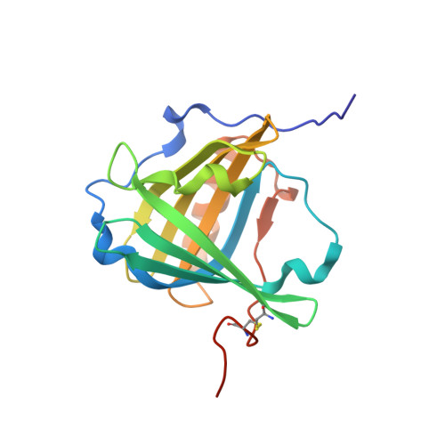
4IAW

Engineered human lipocalin 2 (C26) in complex with Y-DTPA


Experimental Data Snapshot

  • Method: X-RAY DIFFRACTION
  • Resolution: 2.40 Å
  • R-Value Free: 0.270 
  • R-Value Work: 0.224 
  • R-Value Observed: 0.226 

Starting Model: experimental
View more details

wwPDB Validation   3D Report Full Report


Ligand Structure Quality Assessment 


This is version 1.3 of the entry. See complete history


Literature

Structure-guided engineering of Anticalins with improved binding behavior and biochemical characteristics for application in radio-immuno imaging and/or therapy

Eggenstein, E.Eichinger, A.Kim, H.J.Skerra, A.

(2014) J Struct Biol 185: 203-214

  • DOI: https://doi.org/10.1016/j.jsb.2013.03.009
  • Primary Citation of Related Structures:  
    4IAW, 4IAX

  • PubMed Abstract: 

    Modern strategies in radio-immuno therapy and in vivo imaging require robust, small, and specific ligand-binding proteins. In this context we have previously developed artificial lipocalins, so-called Anticalins, with high binding activity toward rare-earth metal-chelate complexes using combinatorial protein design. Here we describe further improvement of the Anticalin C26 via in vitro affinity maturation to yield CL31, which has a fourfold slower dissociation half-life above 2h. Also, we present the crystallographic analyses of both the initial and the improved Anticalin, providing insight into the molecular mechanism of chelated metal binding and the role of amino acid substitutions during the step-wise affinity maturation. Notably, one of the four structurally variable loops that form the ligand pocket in the lipocalin scaffold undergoes a significant conformational change from C26 to CL31, acting as a lid that closes over the accommodated metal-chelate ligand. A systematic mutational study indicated that further improvement of ligand affinity is difficult to achieve while providing clues on the contribution of relevant side chains in the engineered binding pocket. Unexpectedly, some of the amino acid replacements led to strong increases - more then 10-fold - in the yield of soluble protein from periplasmic secretion in Escherichia coli.


  • Organizational Affiliation

    Munich Center for Integrated Protein Science (CIPS-M) and Lehrstuhl für Biologische Chemie, Technische Universität München, Emil-Erlenmeyer-Forum 5, 85350 Freising-Weihenstephan, Germany.


Macromolecules
Find similar proteins by:  (by identity cutoff)  |  3D Structure
Entity ID: 1
MoleculeChains Sequence LengthOrganismDetailsImage
Neutrophil gelatinase-associated lipocalin
A, B, C
188Homo sapiensMutation(s): 23 
Gene Names: HNLLCN2NGAL
UniProt & NIH Common Fund Data Resources
Find proteins for P80188 (Homo sapiens)
Explore P80188 
Go to UniProtKB:  P80188
PHAROS:  P80188
GTEx:  ENSG00000148346 
Entity Groups  
Sequence Clusters30% Identity50% Identity70% Identity90% Identity95% Identity100% Identity
UniProt GroupP80188
Sequence Annotations
Expand
  • Reference Sequence
Small Molecules
Binding Affinity Annotations 
IDSourceBinding Affinity
LIZ PDBBind:  4IAW Kd: 0.23 (nM) from 1 assay(s)
Experimental Data & Validation

Experimental Data

  • Method: X-RAY DIFFRACTION
  • Resolution: 2.40 Å
  • R-Value Free: 0.270 
  • R-Value Work: 0.224 
  • R-Value Observed: 0.226 
  • Space Group: P 41 21 2
Unit Cell:
Length ( Å )Angle ( ˚ )
a = 113.26α = 90
b = 113.26β = 90
c = 119.98γ = 90
Software Package:
Software NamePurpose
MOSFLMdata reduction
SCALAdata scaling
PHASERphasing
REFMACrefinement
PDB_EXTRACTdata extraction
MAR345dtbdata collection

Structure Validation

View Full Validation Report


Ligand Structure Quality Assessment 


Entry History 

Deposition Data

Revision History  (Full details and data files)

  • Version 1.0: 2013-06-19
    Type: Initial release
  • Version 1.1: 2014-02-19
    Changes: Database references
  • Version 1.2: 2023-11-08
    Changes: Data collection, Database references, Derived calculations, Refinement description
  • Version 1.3: 2024-10-30
    Changes: Structure summary