4J09 | pdb_00004j09

Crystal Structure of LpxA bound to RJPXD33


Experimental Data Snapshot

  • Method: X-RAY DIFFRACTION
  • Resolution: 1.90 Å
  • R-Value Free: 
    0.177 (Depositor), 0.180 (DCC) 
  • R-Value Work: 
    0.172 (Depositor), 0.160 (DCC) 
  • R-Value Observed: 
    0.172 (Depositor) 

wwPDB Validation   3D Report Full Report


This is version 1.3 of the entry. See complete history


Literature

Structural Basis for the Recognition of Peptide RJPXD33 by Acyltransferases in Lipid A Biosynthesis.

Jenkins, R.J.Heslip, K.A.Meagher, J.L.Stuckey, J.A.Dotson, G.D.

(2014) J Biological Chem 289: 15527-15535

  • DOI: https://doi.org/10.1074/jbc.M114.564278
  • Primary Citation of Related Structures:  
    4J09

  • PubMed Abstract: 

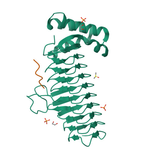
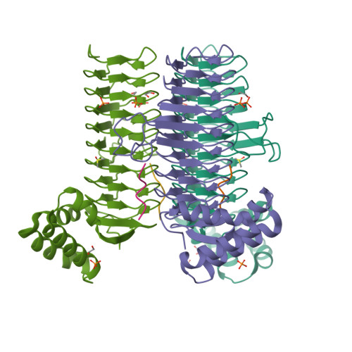
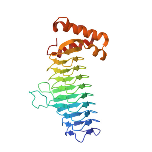
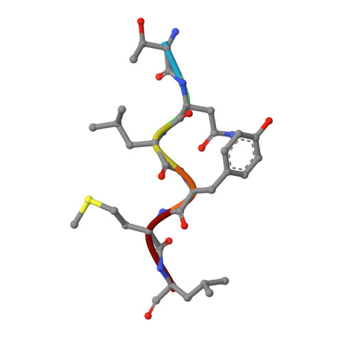
    UDP-N-acetylglucosamine acyltransferase (LpxA) and UDP-3-O-(acyl)-glucosamine acyltransferase (LpxD) constitute the essential, early acyltransferases of lipid A biosynthesis. Recently, an antimicrobial peptide inhibitor, RJPXD33, was identified with dual affinity for LpxA and LpxD. To gain a fundamental understanding of the molecular basis of inhibitor binding, we determined the crystal structure of LpxA from Escherichia coli in complex with RJPXD33 at 1.9 Å resolutions. Our results suggest that the peptide binds in a unique modality that mimics (R)-β-hydroxyacyl pantetheine binding to LpxA and displays how the peptide binds exclusive of the native substrate, acyl-acyl carrier protein. Acyltransferase binding studies with photo-labile RJPXD33 probes and truncations of RJPXD33 validated the structure and provided fundamental insights for future design of small molecule inhibitors. Overlay of the LpxA-RJPXD33 structure with E. coli LpxD identified a complementary peptide binding pocket within LpxD and serves as a model for further biochemical characterization of RJPXD33 binding to LpxD.


  • Organizational Affiliation

    From the Department of Medicinal Chemistry, College of Pharmacy, and.


Macromolecules
Find similar proteins by:  (by identity cutoff)  |  3D Structure
Entity ID: 1
MoleculeChains Sequence LengthOrganismDetailsImage
Acyl-[acyl-carrier-protein]--UDP-N-acetylglucosamine O-acyltransferase262Escherichia coli K-12Mutation(s): 0 
Gene Names: b0181JW0176lpxA
EC: 2.3.1.129
UniProt
Find proteins for P0A722 (Escherichia coli (strain K12))
Explore P0A722 
Go to UniProtKB:  P0A722
Entity Groups  
Sequence Clusters30% Identity50% Identity70% Identity90% Identity95% Identity100% Identity
UniProt GroupP0A722
Sequence Annotations
Expand
  • Reference Sequence

Find similar proteins by:  Sequence   |   3D Structure  

Entity ID: 2
MoleculeChains Sequence LengthOrganismDetailsImage
Putative metabolite transport protein YjhB6Escherichia coli K-12Mutation(s): 0 
UniProt
Find proteins for P39352 (Escherichia coli (strain K12))
Explore P39352 
Go to UniProtKB:  P39352
Entity Groups  
UniProt GroupP39352
Sequence Annotations
Expand
  • Reference Sequence
Experimental Data & Validation

Experimental Data

  • Method: X-RAY DIFFRACTION
  • Resolution: 1.90 Å
  • R-Value Free:  0.177 (Depositor), 0.180 (DCC) 
  • R-Value Work:  0.172 (Depositor), 0.160 (DCC) 
  • R-Value Observed: 0.172 (Depositor) 
Space Group: P 21 3
Unit Cell:
Length ( Å )Angle ( ˚ )
a = 96.399α = 90
b = 96.399β = 90
c = 96.399γ = 90
Software Package:
Software NamePurpose
PHASERphasing
BUSTER-TNTrefinement
PDB_EXTRACTdata extraction
HKL-2000data collection
HKL-2000data reduction
HKL-2000data scaling
BUSTERrefinement

Structure Validation

View Full Validation Report


Entry History 

Deposition Data

Revision History  (Full details and data files)

  • Version 1.0: 2014-04-23
    Type: Initial release
  • Version 1.1: 2014-06-11
    Changes: Database references
  • Version 1.2: 2014-06-18
    Changes: Database references
  • Version 1.3: 2024-02-28
    Changes: Data collection, Database references, Derived calculations