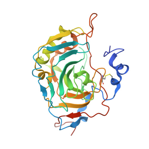
4LU3

The crystal structure of the human carbonic anhydrase XIV


Experimental Data Snapshot

  • Method: X-RAY DIFFRACTION
  • Resolution: 2.00 Å
  • R-Value Free: 0.206 
  • R-Value Work: 0.181 
  • R-Value Observed: 0.181 

Starting Model: experimental
View more details

wwPDB Validation   3D Report Full Report


Ligand Structure Quality Assessment 


This is version 2.1 of the entry. See complete history


Literature

The structural comparison between membrane-associated human carbonic anhydrases provides insights into drug design of selective inhibitors.

Alterio, V.Pan, P.Parkkila, S.Buonanno, M.Supuran, C.T.Monti, S.M.De Simone, G.

(2014) Biopolymers 101: 769-778

  • DOI: https://doi.org/10.1002/bip.22456
  • Primary Citation of Related Structures:  
    4LU3

  • PubMed Abstract: 

    Carbonic anhydrase isoform XIV (CA XIV) is the last member of the human (h) CA family discovered so far, being localized in brain, kidneys, colon, small intestine, urinary bladder, liver, and spinal cord. It has recently been described as a possible drug target for treatment of epilepsy, some retinopathies as well as some skin tumors. Human carbonic anhydrase (hCA) XIV is a membrane-associated protein consisting of an N-terminal extracellular domain, a putative transmembrane region, and a small cytoplasmic tail. In this article, we report the expression, purification, and the crystallographic structure of the entire extracellular domain of this enzyme. The analysis of the structure revealed the typical α-CA fold, in which a 10-stranded β-sheet forms the core of the molecule, while the comparison with all the other membrane associated isoforms (hCAs IV, IX, and XII) allowed to identify the diverse oligomeric arrangement and the sequence and structural differences observed in the region 127-136 as the main factors to consider in the design of selective inhibitors for each one of the membrane associated α-CAs.


  • Organizational Affiliation

    Institute of Biostructures and Bioimaging, CNR, Via Mezzocannone 16, 80134, Naples, Italy.


Macromolecules
Find similar proteins by:  (by identity cutoff)  |  3D Structure
Entity ID: 1
MoleculeChains Sequence LengthOrganismDetailsImage
Carbonic anhydrase 14279Homo sapiensMutation(s): 0 
Gene Names: CA14UNQ690/PRO1335
EC: 4.2.1.1
UniProt & NIH Common Fund Data Resources
Find proteins for Q9ULX7 (Homo sapiens)
Explore Q9ULX7 
Go to UniProtKB:  Q9ULX7
PHAROS:  Q9ULX7
GTEx:  ENSG00000118298 
Entity Groups  
Sequence Clusters30% Identity50% Identity70% Identity90% Identity95% Identity100% Identity
UniProt GroupQ9ULX7
Glycosylation
Glycosylation Sites: 1Go to GlyGen: Q9ULX7-1
Sequence Annotations
Expand
  • Reference Sequence
Oligosaccharides

Help

Entity ID: 2
MoleculeChains Length2D Diagram Glycosylation3D Interactions
alpha-D-mannopyranose-(1-3)-[alpha-D-mannopyranose-(1-6)]beta-D-mannopyranose-(1-4)-2-acetamido-2-deoxy-beta-D-glucopyranose-(1-4)-2-acetamido-2-deoxy-beta-D-glucopyranose
B
5N-Glycosylation
Glycosylation Resources
GlyTouCan:  G22768VO
GlyCosmos:  G22768VO
GlyGen:  G22768VO
Small Molecules
Ligands 4 Unique
IDChains Name / Formula / InChI Key2D Diagram3D Interactions
AZM
Query on AZM

Download Ideal Coordinates CCD File 
D [auth A]5-ACETAMIDO-1,3,4-THIADIAZOLE-2-SULFONAMIDE
C4 H6 N4 O3 S2
BZKPWHYZMXOIDC-UHFFFAOYSA-N
SO4
Query on SO4

Download Ideal Coordinates CCD File 
I [auth A],
J [auth A]
SULFATE ION
O4 S
QAOWNCQODCNURD-UHFFFAOYSA-L
GOL
Query on GOL

Download Ideal Coordinates CCD File 
E [auth A],
F [auth A],
G [auth A],
H [auth A]
GLYCEROL
C3 H8 O3
PEDCQBHIVMGVHV-UHFFFAOYSA-N
ZN
Query on ZN

Download Ideal Coordinates CCD File 
C [auth A]ZINC ION
Zn
PTFCDOFLOPIGGS-UHFFFAOYSA-N
Experimental Data & Validation

Experimental Data

  • Method: X-RAY DIFFRACTION
  • Resolution: 2.00 Å
  • R-Value Free: 0.206 
  • R-Value Work: 0.181 
  • R-Value Observed: 0.181 
  • Space Group: P 41 21 2
Unit Cell:
Length ( Å )Angle ( ˚ )
a = 88.61α = 90
b = 88.61β = 90
c = 108.9γ = 90
Software Package:
Software NamePurpose
CrystalCleardata collection
AMoREphasing
CNSrefinement
HKL-2000data reduction
HKL-2000data scaling

Structure Validation

View Full Validation Report


Ligand Structure Quality Assessment 


Entry History 

Deposition Data

Revision History  (Full details and data files)

  • Version 1.0: 2014-01-01
    Type: Initial release
  • Version 1.1: 2014-01-08
    Changes: Other
  • Version 1.2: 2014-05-14
    Changes: Database references
  • Version 2.0: 2020-07-29
    Type: Remediation
    Reason: Carbohydrate remediation
    Changes: Atomic model, Data collection, Database references, Derived calculations, Structure summary
  • Version 2.1: 2023-09-20
    Changes: Data collection, Database references, Refinement description, Structure summary