4RLT

Crystal Structure of (3R)-hydroxyacyl-ACP dehydratase HadAB hetero-dimer from Mycobacterium tuberculosis complexed with Fisetin


Experimental Data Snapshot

  • Method: X-RAY DIFFRACTION
  • Resolution: 2.05 Å
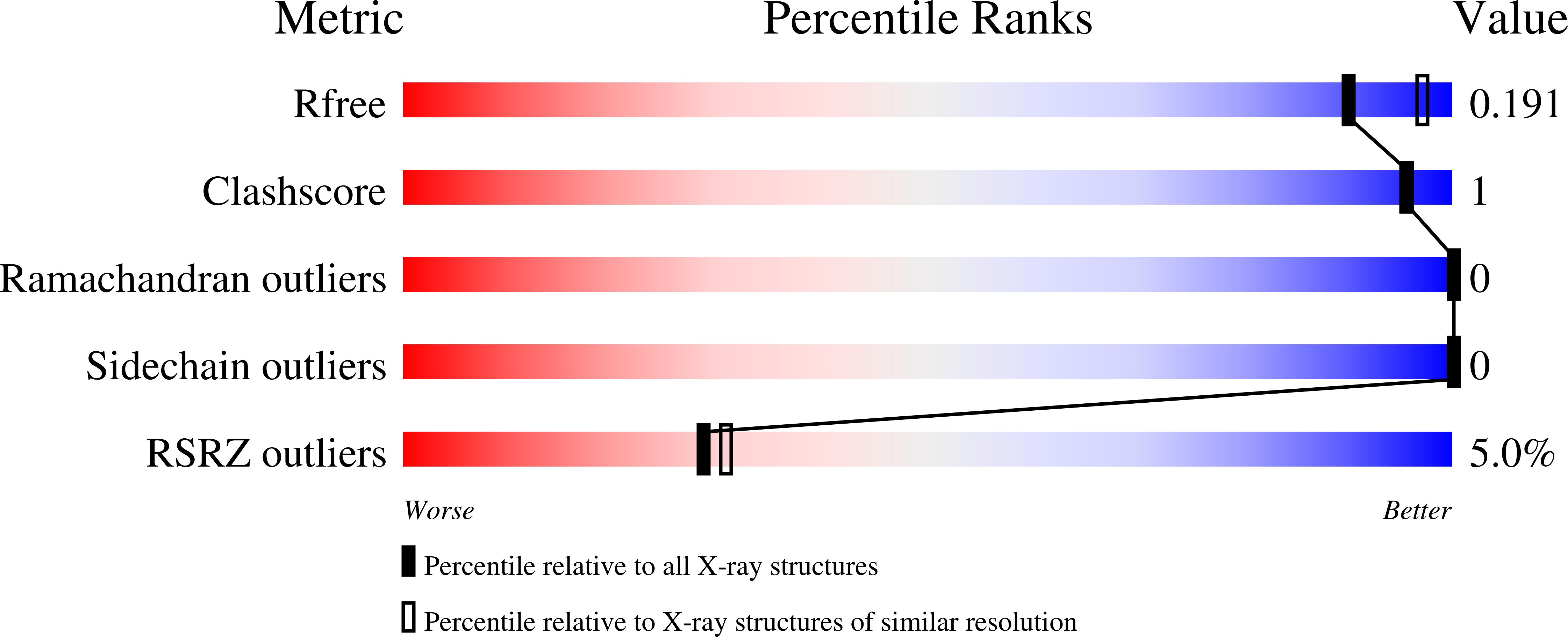
  • R-Value Free: 0.190 
  • R-Value Work: 0.158 
  • R-Value Observed: 0.159 

Starting Model: experimental
View more details

wwPDB Validation   3D Report Full Report


Ligand Structure Quality Assessment 


This is version 1.2 of the entry. See complete history


Literature

Molecular basis for the inhibition of beta-hydroxyacyl-ACP dehydratase HadAB complex from Mycobacterium tuberculosis by flavonoid inhibitors.

Dong, Y.Qiu, X.Shaw, N.Xu, Y.Sun, Y.Li, X.Li, J.Rao, Z.

(2015) Protein Cell 6: 504-517

  • DOI: https://doi.org/10.1007/s13238-015-0181-1
  • Primary Citation of Related Structures:  
    4RLJ, 4RLT, 4RLU, 4RLW

  • PubMed Abstract: 

    Dehydration is one of the key steps in the biosynthesis of mycolic acids and is vital to the growth of Mycobacterium tuberculosis (Mtb). Consequently, stalling dehydration cures tuberculosis (TB). Clinically used anti-TB drugs like thiacetazone (TAC) and isoxyl (ISO) as well as flavonoids inhibit the enzyme activity of the β-hydroxyacyl-ACP dehydratase HadAB complex. How this inhibition is exerted, has remained an enigma for years. Here, we describe the first crystal structures of the MtbHadAB complex bound with flavonoid inhibitor butein, 2',4,4'-trihydroxychalcone or fisetin. Despite sharing no sequence identity from Blast, HadA and HadB adopt a very similar hotdog fold. HadA forms a tight dimer with HadB in which the proteins are sitting side-by-side, but are oriented anti-parallel. While HadB contributes the catalytically critical His-Asp dyad, HadA binds the fatty acid substrate in a long channel. The atypical double hotdog fold with a single active site formed by MtbHadAB gives rise to a long, narrow cavity that vertically traverses the fatty acid binding channel. At the base of this cavity lies Cys61, which upon mutation to Ser confers drug-resistance in TB patients. We show that inhibitors bind in this cavity and protrude into the substrate binding channel. Thus, inhibitors of MtbHadAB exert their effect by occluding substrate from the active site. The unveiling of this mechanism of inhibition paves the way for accelerating development of next generation of anti-TB drugs.


  • Organizational Affiliation

    National Laboratory of Biomacromolecules, Institute of Biophysics, Chinese Academy of Sciences, Beijing, 100101, China.


Macromolecules
Find similar proteins by:  (by identity cutoff)  |  3D Structure
Entity ID: 1
MoleculeChains Sequence LengthOrganismDetailsImage
(3R)-hydroxyacyl-ACP dehydratase subunit HadA158Mycobacterium tuberculosis H37RvMutation(s): 0 
Gene Names: hadAP425_00663RVBD_0635
EC: 4.2.1.59
UniProt
Find proteins for P9WFK1 (Mycobacterium tuberculosis (strain ATCC 25618 / H37Rv))
Explore P9WFK1 
Go to UniProtKB:  P9WFK1
Entity Groups  
Sequence Clusters30% Identity50% Identity70% Identity90% Identity95% Identity100% Identity
UniProt GroupP9WFK1
Sequence Annotations
Expand
  • Reference Sequence
Find similar proteins by:  (by identity cutoff)  |  3D Structure
Entity ID: 2
MoleculeChains Sequence LengthOrganismDetailsImage
(3R)-hydroxyacyl-ACP dehydratase subunit HadB147Mycobacterium tuberculosis H37RvMutation(s): 0 
Gene Names: hadBP425_00664Rv0636RVBD_0636
EC: 4.2.1.59
UniProt
Find proteins for I6WYY7 (Mycobacterium tuberculosis (strain ATCC 25618 / H37Rv))
Explore I6WYY7 
Go to UniProtKB:  I6WYY7
Entity Groups  
Sequence Clusters30% Identity50% Identity70% Identity90% Identity95% Identity100% Identity
UniProt GroupI6WYY7
Sequence Annotations
Expand
  • Reference Sequence
Experimental Data & Validation

Experimental Data

  • Method: X-RAY DIFFRACTION
  • Resolution: 2.05 Å
  • R-Value Free: 0.190 
  • R-Value Work: 0.158 
  • R-Value Observed: 0.159 
  • Space Group: P 41 21 2
Unit Cell:
Length ( Å )Angle ( ˚ )
a = 82.258α = 90
b = 82.258β = 90
c = 140.161γ = 90
Software Package:
Software NamePurpose
DENZOdata reduction
SCALEPACKdata scaling
PHENIXrefinement
PDB_EXTRACTdata extraction
HKL-2000data collection
HKL-2000data reduction
HKL-2000data scaling
PHASERphasing

Structure Validation

View Full Validation Report


Ligand Structure Quality Assessment 


Entry History 

Deposition Data

Revision History  (Full details and data files)

  • Version 1.0: 2015-10-21
    Type: Initial release
  • Version 1.1: 2017-11-22
    Changes: Refinement description
  • Version 1.2: 2023-09-20
    Changes: Data collection, Database references, Derived calculations, Refinement description, Structure summary