4Z6V

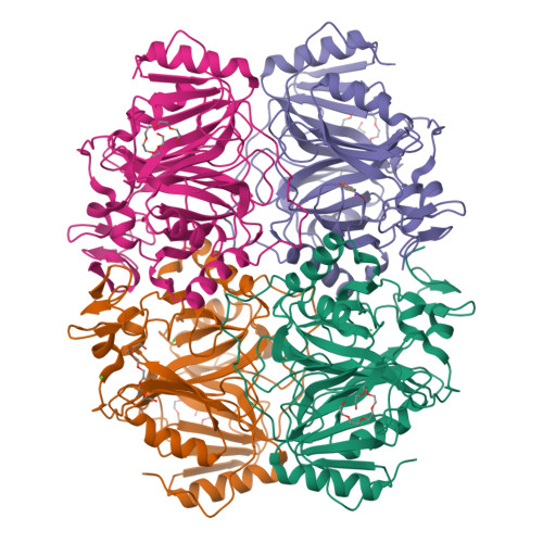
Structure of H200Q variant of Homoprotocatechuate 2,3-Dioxygenase from B.fuscum in complex with 4-nitrocatechol at 1.37 Ang resolution

  • Classification: OXIDOREDUCTASE
  • Organism(s): Brevibacterium fuscum
  • Expression System: Escherichia coli
  • Mutation(s): Yes 

  • Deposited: 2015-04-06 Released: 2015-08-26 
  • Deposition Author(s): Kovaleva, E.G., Lipscomb, J.D.
  • Funding Organization(s): Biotechnology and Biological Sciences Research Council (BBSRC), National Institutes of Health/National Institute of General Medical Sciences (NIH/NIGMS)

Experimental Data Snapshot

  • Method: X-RAY DIFFRACTION
  • Resolution: 1.37 Å
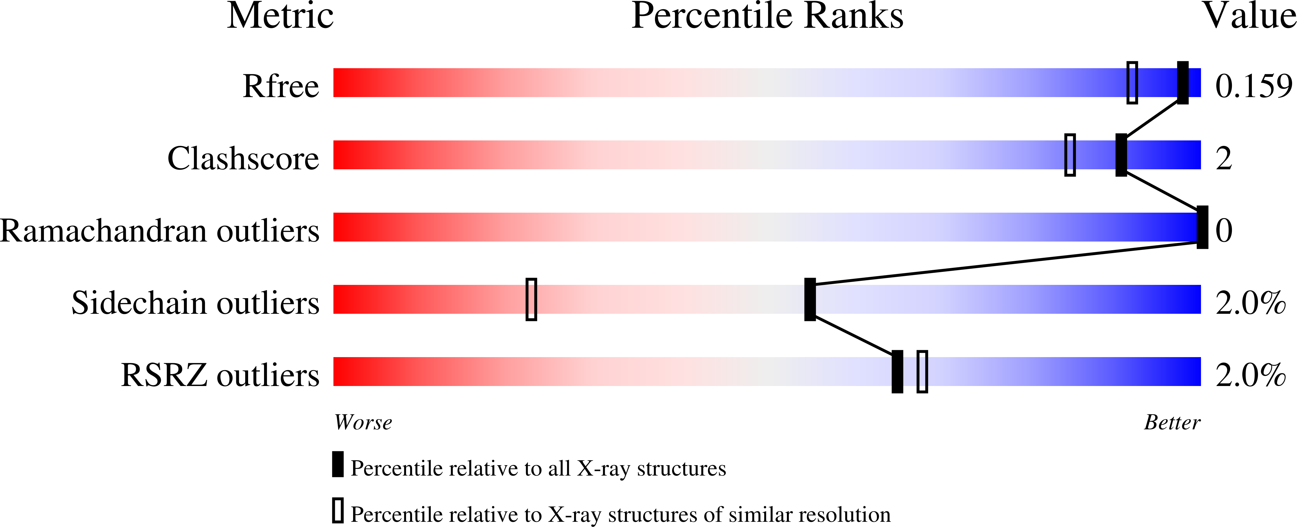
  • R-Value Free: 0.157 
  • R-Value Work: 0.119 
  • R-Value Observed: 0.121 

Starting Model: experimental
View more details

wwPDB Validation   3D Report Full Report


Ligand Structure Quality Assessment 

Created with Raphaël 2.3.0Worse 01 BetterLigand structure goodness of fit to experimental dataBest fitted 4NCClick on this verticalbar to view details

This is version 1.5 of the entry. See complete history


Literature

Structural Basis for Substrate and Oxygen Activation in Homoprotocatechuate 2,3-Dioxygenase: Roles of Conserved Active Site Histidine 200.

Kovaleva, E.G.Rogers, M.S.Lipscomb, J.D.

(2015) Biochemistry 54: 5329-5339

  • DOI: https://doi.org/10.1021/acs.biochem.5b00709
  • Primary Citation of Related Structures:  
    4Z6L, 4Z6M, 4Z6N, 4Z6O, 4Z6P, 4Z6Q, 4Z6R, 4Z6S, 4Z6T, 4Z6U, 4Z6V, 4Z6W, 4Z6Z

  • PubMed Abstract: 

    Kinetic and spectroscopic studies have shown that the conserved active site residue His200 of the extradiol ring-cleaving homoprotocatechuate 2,3-dioxygenase (FeHPCD) from Brevibacterium fuscum is critical for efficient catalysis. The roles played by this residue are probed here by analysis of the steady-state kinetics, pH dependence, and X-ray crystal structures of the FeHPCD position 200 variants His200Asn, His200Gln, and His200Glu alone and in complex with three catecholic substrates (homoprotocatechuate, 4-sulfonylcatechol, and 4-nitrocatechol) possessing substituents with different inductive capacity. Structures determined at 1.35-1.75 Å resolution show that there is essentially no change in overall active site architecture or substrate binding mode for these variants when compared to the structures of the wild-type enzyme and its analogous complexes. This shows that the maximal 50-fold decrease in kcat for ring cleavage, the dramatic changes in pH dependence, and the switch from ring cleavage to ring oxidation of 4-nitrocatechol by the FeHPCD variants can be attributed specifically to the properties of the altered second-sphere residue and the substrate. The results suggest that proton transfer is necessary for catalysis, and that it occurs most efficiently when the substrate provides the proton and His200 serves as a catalyst. However, in the absence of an available substrate proton, a defined proton-transfer pathway in the protein can be utilized. Changes in the steric bulk and charge of the residue at position 200 appear to be capable of altering the rate-limiting step in catalysis and, perhaps, the nature of the reactive species.


  • Organizational Affiliation

    Institute of Molecular and Cellular Biology, University of Leeds , Leeds LS2 9JT, U.K.


Macromolecules
Find similar proteins by:  (by identity cutoff)  |  3D Structure
Entity ID: 1
MoleculeChains Sequence LengthOrganismDetailsImage
Homoprotocatechuate 2,3-dioxygenase
A, B, C, D
365Brevibacterium fuscumMutation(s): 1 
UniProt
Find proteins for Q45135 (Brevibacterium fuscum)
Explore Q45135 
Go to UniProtKB:  Q45135
Entity Groups  
Sequence Clusters30% Identity50% Identity70% Identity90% Identity95% Identity100% Identity
UniProt GroupQ45135
Sequence Annotations
Expand
  • Reference Sequence
Small Molecules
Ligands 5 Unique
IDChains Name / Formula / InChI Key2D Diagram3D Interactions
P6G
Query on P6G

Download Ideal Coordinates CCD File 
F [auth A],
J [auth B],
K [auth B],
N [auth C],
Q [auth D]
HEXAETHYLENE GLYCOL
C12 H26 O7
IIRDTKBZINWQAW-UHFFFAOYSA-N
4NC
Query on 4NC

Download Ideal Coordinates CCD File 
O [auth C]4-NITROCATECHOL
C6 H5 N O4
XJNPNXSISMKQEX-UHFFFAOYSA-N
FE2
Query on FE2

Download Ideal Coordinates CCD File 
E [auth A],
H [auth B],
M [auth C],
P [auth D]
FE (II) ION
Fe
CWYNVVGOOAEACU-UHFFFAOYSA-N
CA
Query on CA

Download Ideal Coordinates CCD File 
I [auth B]CALCIUM ION
Ca
BHPQYMZQTOCNFJ-UHFFFAOYSA-N
CL
Query on CL

Download Ideal Coordinates CCD File 
G [auth A],
L [auth B],
R [auth D]
CHLORIDE ION
Cl
VEXZGXHMUGYJMC-UHFFFAOYSA-M
Experimental Data & Validation

Experimental Data

  • Method: X-RAY DIFFRACTION
  • Resolution: 1.37 Å
  • R-Value Free: 0.157 
  • R-Value Work: 0.119 
  • R-Value Observed: 0.121 
  • Space Group: P 21 21 2
Unit Cell:
Length ( Å )Angle ( ˚ )
a = 110.18α = 90
b = 150.635β = 90
c = 96.103γ = 90
Software Package:
Software NamePurpose
REFMACrefinement
SCALAdata scaling
PDB_EXTRACTdata extraction
XDSdata reduction
MOLREPphasing

Structure Validation

View Full Validation Report


Ligand Structure Quality Assessment 

Created with Raphaël 2.3.0Worse 01 BetterLigand structure goodness of fit to experimental dataBest fitted 4NCClick on this verticalbar to view details

Entry History & Funding Information

Deposition Data


Funding OrganizationLocationGrant Number
Biotechnology and Biological Sciences Research Council (BBSRC)United KingdomBB/H001905/1
National Institutes of Health/National Institute of General Medical Sciences (NIH/NIGMS)United StatesGM 24689

Revision History  (Full details and data files)

  • Version 1.0: 2015-08-26
    Type: Initial release
  • Version 1.1: 2015-09-16
    Changes: Database references
  • Version 1.2: 2017-09-20
    Changes: Author supporting evidence, Database references, Derived calculations, Structure summary
  • Version 1.3: 2019-12-25
    Changes: Author supporting evidence
  • Version 1.4: 2020-01-01
    Changes: Author supporting evidence
  • Version 1.5: 2023-09-27
    Changes: Data collection, Database references, Derived calculations, Refinement description