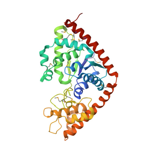
5DK4

Crystal structure analysis of Tryptophanyl-trna synthetase from Bacillus stearothermophilus in complex with indolmycin and Mg*ATP


Experimental Data Snapshot

  • Method: X-RAY DIFFRACTION
  • Resolution: 1.90 Å
  • R-Value Free: 0.189 
  • R-Value Work: 0.169 
  • R-Value Observed: 0.170 

wwPDB Validation   3D Report Full Report


Ligand Structure Quality Assessment 


This is version 1.5 of the entry. See complete history


Literature

Selective Inhibition of Bacterial Tryptophanyl-tRNA Synthetases by Indolmycin Is Mechanism-based.

Williams, T.L.Yin, Y.W.Carter, C.W.

(2016) J Biol Chem 291: 255-265

  • DOI: https://doi.org/10.1074/jbc.M115.690321
  • Primary Citation of Related Structures:  
    5DK4

  • PubMed Abstract: 

    Indolmycin is a natural tryptophan analog that competes with tryptophan for binding to tryptophanyl-tRNA synthetase (TrpRS) enzymes. Bacterial and eukaryotic cytosolic TrpRSs have comparable affinities for tryptophan (Km ∼ 2 μm), and yet only bacterial TrpRSs are inhibited by indolmycin. Despite the similarity between these ligands, Bacillus stearothermophilus (Bs)TrpRS preferentially binds indolmycin ∼1500-fold more tightly than its tryptophan substrate. Kinetic characterization and crystallographic analysis of BsTrpRS allowed us to probe novel aspects of indolmycin inhibitory action. Previous work had revealed that long range coupling to residues within an allosteric region called the D1 switch of BsTrpRS positions the Mg(2+) ion in a manner that allows it to assist in transition state stabilization. The Mg(2+) ion in the inhibited complex forms significantly closer contacts with non-bridging oxygen atoms from each phosphate group of ATP and three water molecules than occur in the (presumably catalytically competent) pre-transition state (preTS) crystal structures. We propose that this altered coordination stabilizes a ground state Mg(2+)·ATP configuration, accounting for the high affinity inhibition of BsTrpRS by indolmycin. Conversely, both the ATP configuration and Mg(2+) coordination in the human cytosolic (Hc)TrpRS preTS structure differ greatly from the BsTrpRS preTS structure. The effect of these differences is that catalysis occurs via a different transition state stabilization mechanism in HcTrpRS with a yet-to-be determined role for Mg(2+). Modeling indolmycin into the tryptophan binding site points to steric hindrance and an inability to retain the interactions used for tryptophan substrate recognition as causes for the 1000-fold weaker indolmycin affinity to HcTrpRS.


  • Organizational Affiliation

    From the Department of Biochemistry and Biophysics, University of North Carolina, Chapel Hill, North Carolina 27599-7260 and tishan.williams@gmail.com.


Macromolecules
Find similar proteins by:  (by identity cutoff)  |  3D Structure
Entity ID: 1
MoleculeChains Sequence LengthOrganismDetailsImage
Tryptophan--tRNA ligase329Geobacillus stearothermophilusMutation(s): 0 
Gene Names: trpS
EC: 6.1.1.2
UniProt
Find proteins for P00953 (Geobacillus stearothermophilus)
Explore P00953 
Go to UniProtKB:  P00953
Entity Groups  
Sequence Clusters30% Identity50% Identity70% Identity90% Identity95% Identity100% Identity
UniProt GroupP00953
Sequence Annotations
Expand
  • Reference Sequence
Small Molecules
Ligands 4 Unique
IDChains Name / Formula / InChI Key2D Diagram3D Interactions
ATP
Query on ATP

Download Ideal Coordinates CCD File 
B [auth A]ADENOSINE-5'-TRIPHOSPHATE
C10 H16 N5 O13 P3
ZKHQWZAMYRWXGA-KQYNXXCUSA-N
5BX
Query on 5BX

Download Ideal Coordinates CCD File 
E [auth A](5S)-5-[(1R)-1-(1H-indol-3-yl)ethyl]-2-(methylamino)-1,3-oxazol-4(5H)-one
C14 H15 N3 O2
GNTVWGDQPXCYBV-PELKAZGASA-N
GOL
Query on GOL

Download Ideal Coordinates CCD File 
D [auth A]GLYCEROL
C3 H8 O3
PEDCQBHIVMGVHV-UHFFFAOYSA-N
MG
Query on MG

Download Ideal Coordinates CCD File 
C [auth A]MAGNESIUM ION
Mg
JLVVSXFLKOJNIY-UHFFFAOYSA-N
Modified Residues  1 Unique
IDChains TypeFormula2D DiagramParent
MSE
Query on MSE
A
L-PEPTIDE LINKINGC5 H11 N O2 SeMET
Experimental Data & Validation

Experimental Data

  • Method: X-RAY DIFFRACTION
  • Resolution: 1.90 Å
  • R-Value Free: 0.189 
  • R-Value Work: 0.169 
  • R-Value Observed: 0.170 
  • Space Group: P 43 21 2
Unit Cell:
Length ( Å )Angle ( ˚ )
a = 62.041α = 90
b = 62.041β = 90
c = 219.058γ = 90
Software Package:
Software NamePurpose
PHENIXrefinement
XDSdata reduction
SCALAdata scaling
PHENIXphasing

Structure Validation

View Full Validation Report


Ligand Structure Quality Assessment 


Entry History & Funding Information

Deposition Data


Funding OrganizationLocationGrant Number
National Institutes of Health/National Institute of General Medical Sciences (NIH/NIGMS)United StatesGM40906

Revision History  (Full details and data files)

  • Version 1.0: 2015-11-18
    Type: Initial release
  • Version 1.1: 2015-11-25
    Changes: Database references
  • Version 1.2: 2016-01-13
    Changes: Database references
  • Version 1.3: 2017-09-20
    Changes: Author supporting evidence, Database references, Derived calculations
  • Version 1.4: 2019-12-25
    Changes: Author supporting evidence
  • Version 1.5: 2024-10-23
    Changes: Data collection, Database references, Derived calculations, Structure summary