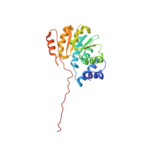
5FHQ

Crystal structure of (WT) Rat Catechol-O-Methyltransferase in complex with AdoMet and 3,5-dinitrocatechol (DNC)


Experimental Data Snapshot

  • Method: X-RAY DIFFRACTION
  • Resolution: 1.63 Å
  • R-Value Free: 0.188 
  • R-Value Work: 0.155 
  • R-Value Observed: 0.156 

Starting Model: experimental
View more details

wwPDB Validation   3D Report Full Report


Ligand Structure Quality Assessment 


This is version 2.0 of the entry. See complete history


Literature

Effects of Active-Site Modification and Quaternary Structure on the Regioselectivity of Catechol-O-Methyltransferase.

Law, B.J.Bennett, M.R.Thompson, M.L.Levy, C.Shepherd, S.A.Leys, D.Micklefield, J.

(2016) Angew Chem Int Ed Engl 55: 2683-2687

  • DOI: https://doi.org/10.1002/anie.201508287
  • Primary Citation of Related Structures:  
    5FHQ, 5FHR

  • PubMed Abstract: 

    Catechol-O-methyltransferase (COMT), an important therapeutic target in the treatment of Parkinson's disease, is also being developed for biocatalytic processes, including vanillin production, although lack of regioselectivity has precluded its more widespread application. By using structural and mechanistic information, regiocomplementary COMT variants were engineered that deliver either meta- or para-methylated catechols. X-ray crystallography further revealed how the active-site residues and quaternary structure govern regioselectivity. Finally, analogues of AdoMet are accepted by the regiocomplementary COMT mutants and can be used to prepare alkylated catechols, including ethyl vanillin.


  • Organizational Affiliation

    School of Chemistry & Manchester Institute of Biotechnology, University of Manchester, 131 Princess Street, Manchester, M1 7DN, UK.


Macromolecules
Find similar proteins by:  (by identity cutoff)  |  3D Structure
Entity ID: 1
MoleculeChains Sequence LengthOrganismDetailsImage
Catechol O-methyltransferase213Rattus norvegicusMutation(s): 0 
Gene Names: Comt
EC: 2.1.1.6
UniProt
Find proteins for P22734 (Rattus norvegicus)
Explore P22734 
Go to UniProtKB:  P22734
Entity Groups  
Sequence Clusters30% Identity50% Identity70% Identity90% Identity95% Identity100% Identity
UniProt GroupP22734
Sequence Annotations
Expand
  • Reference Sequence
Small Molecules
Ligands 3 Unique
IDChains Name / Formula / InChI Key2D Diagram3D Interactions
SAM
Query on SAM

Download Ideal Coordinates CCD File 
B [auth A]S-ADENOSYLMETHIONINE
C15 H22 N6 O5 S
MEFKEPWMEQBLKI-FCKMPRQPSA-N
DNC
Query on DNC

Download Ideal Coordinates CCD File 
C [auth A]3,5-DINITROCATECHOL
C6 H4 N2 O6
VDCDWNDTNSWDFJ-UHFFFAOYSA-N
MG
Query on MG

Download Ideal Coordinates CCD File 
D [auth A]MAGNESIUM ION
Mg
JLVVSXFLKOJNIY-UHFFFAOYSA-N
Experimental Data & Validation

Experimental Data

  • Method: X-RAY DIFFRACTION
  • Resolution: 1.63 Å
  • R-Value Free: 0.188 
  • R-Value Work: 0.155 
  • R-Value Observed: 0.156 
  • Space Group: P 32 2 1
Unit Cell:
Length ( Å )Angle ( ˚ )
a = 50.72α = 90
b = 50.72β = 90
c = 167.55γ = 120
Software Package:
Software NamePurpose
PHENIXrefinement
xia2data reduction
xia2data scaling
PHASERphasing

Structure Validation

View Full Validation Report


Ligand Structure Quality Assessment 


Entry History 

Deposition Data

  • Released Date: 2016-02-03 
  • Deposition Author(s): Levy, C.

Revision History  (Full details and data files)

  • Version 1.0: 2016-02-03
    Type: Initial release
  • Version 1.1: 2016-02-24
    Changes: Database references
  • Version 2.0: 2024-01-10
    Changes: Atomic model, Data collection, Database references, Refinement description