5HG1 | pdb_00005hg1

Crystal Structure of Human Hexokinase 2 with cmpd 1, a C-2-substituted glucosamine


Experimental Data Snapshot

  • Method: X-RAY DIFFRACTION
  • Resolution: 2.76 Å
  • R-Value Free: 
    0.266 (Depositor), 0.270 (DCC) 
  • R-Value Work: 
    0.218 (Depositor), 0.220 (DCC) 
  • R-Value Observed: 
    0.220 (Depositor) 

wwPDB Validation   3D Report Full Report


Ligand Structure Quality Assessment 

Created with Raphaël 2.3.0Worse 01 BetterLigand structure goodness of fit to experimental dataBest fitted 62CClick on this verticalbar to view detailsBest fitted BG6Click on this verticalbar to view details

This is version 1.2 of the entry. See complete history


Literature

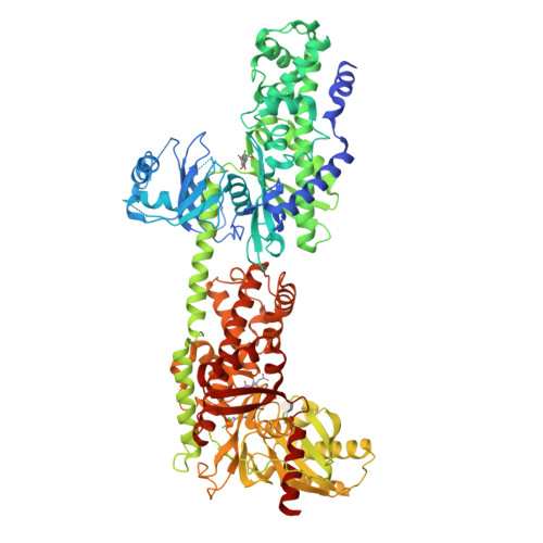
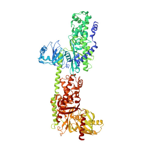
Discovery of a Novel 2,6-Disubstituted Glucosamine Series of Potent and Selective Hexokinase 2 Inhibitors.

Lin, H.Zeng, J.Xie, R.Schulz, M.J.Tedesco, R.Qu, J.Erhard, K.F.Mack, J.F.Raha, K.Rendina, A.R.Szewczuk, L.M.Kratz, P.M.Jurewicz, A.J.Cecconie, T.Martens, S.McDevitt, P.J.Martin, J.D.Chen, S.B.Jiang, Y.Nickels, L.Schwartz, B.J.Smallwood, A.Zhao, B.Campobasso, N.Qian, Y.Briand, J.Rominger, C.M.Oleykowski, C.Hardwicke, M.A.Luengo, J.I.

(2016) ACS Med Chem Lett 7: 217-222

  • DOI: https://doi.org/10.1021/acsmedchemlett.5b00214
  • Primary Citation of Related Structures:  
    5HEX, 5HFU, 5HG1

  • PubMed Abstract: 

    A novel series of potent and selective hexokinase 2 (HK2) inhibitors, 2,6-disubstituted glucosamines, has been identified based on HTS hits, exemplified by compound 1. Inhibitor-bound crystal structures revealed that the HK2 enzyme could adopt an "induced-fit" conformation. The SAR study led to the identification of potent HK2 inhibitors, such as compound 34 with greater than 100-fold selectivity over HK1. Compound 25 inhibits in situ glycolysis in a UM-UC-3 bladder tumor cell line via (13)CNMR measurement of [3-(13)C]lactate produced from [1,6-(13)C2]glucose added to the cell culture.


  • Organizational Affiliation

    Cancer Metabolism Chemistry; Cancer Metabolism Biology; and Platform Technology & Sciences, GlaxoSmithKline , 1250 South Collegeville Road, Collegeville, Pennsylvania 19426-0989, United States.


Macromolecules
Find similar proteins by:  (by identity cutoff)  |  3D Structure
Entity ID: 1
MoleculeChains Sequence LengthOrganismDetailsImage
Hexokinase-2922Homo sapiensMutation(s): 0 
Gene Names: HK2
EC: 2.7.1.1
UniProt & NIH Common Fund Data Resources
Find proteins for P52789 (Homo sapiens)
Explore P52789 
Go to UniProtKB:  P52789
PHAROS:  P52789
GTEx:  ENSG00000159399 
Entity Groups  
Sequence Clusters30% Identity50% Identity70% Identity90% Identity95% Identity100% Identity
UniProt GroupP52789
Sequence Annotations
Expand
  • Reference Sequence
Experimental Data & Validation

Experimental Data

  • Method: X-RAY DIFFRACTION
  • Resolution: 2.76 Å
  • R-Value Free:  0.266 (Depositor), 0.270 (DCC) 
  • R-Value Work:  0.218 (Depositor), 0.220 (DCC) 
  • R-Value Observed: 0.220 (Depositor) 
Space Group: P 31 2 1
Unit Cell:
Length ( Å )Angle ( ˚ )
a = 165.12α = 90
b = 165.12β = 90
c = 126.478γ = 120
Software Package:
Software NamePurpose
PHENIXrefinement
XDSdata reduction
Aimlessdata scaling
PHENIXphasing

Structure Validation

View Full Validation Report


Ligand Structure Quality Assessment 

Created with Raphaël 2.3.0Worse 01 BetterLigand structure goodness of fit to experimental dataBest fitted 62CClick on this verticalbar to view detailsBest fitted BG6Click on this verticalbar to view details

Entry History 

Deposition Data

Revision History  (Full details and data files)

  • Version 1.0: 2016-03-30
    Type: Initial release
  • Version 1.1: 2020-07-29
    Type: Remediation
    Reason: Carbohydrate remediation
    Changes: Data collection, Derived calculations, Structure summary
  • Version 1.2: 2024-03-06
    Changes: Data collection, Database references, Structure summary