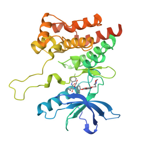
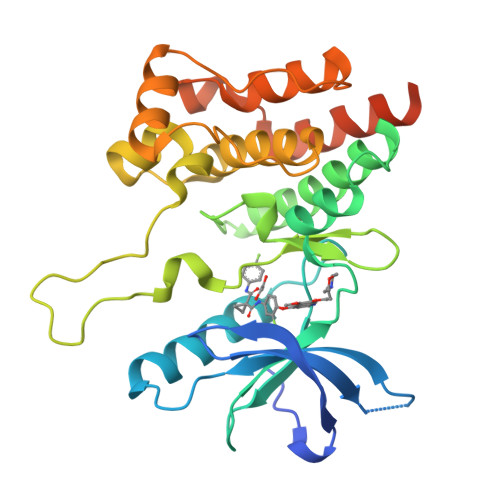
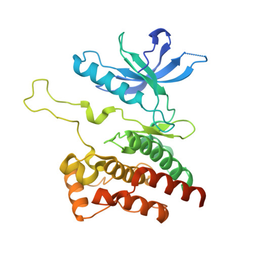
5IA4

Crystal Structure of Ephrin A2 (EphA2) Receptor Protein Kinase with foretinib (XL880)


Experimental Data Snapshot

  • Method: X-RAY DIFFRACTION
  • Resolution: 1.80 Å
  • R-Value Free: 0.226 
  • R-Value Work: 0.189 
  • R-Value Observed: 0.190 

Starting Model: experimental
View more details

wwPDB Validation   3D Report Full Report


Ligand Structure Quality Assessment 


This is version 1.6 of the entry. See complete history


Literature

Chemical Proteomics and Structural Biology Define EPHA2 Inhibition by Clinical Kinase Drugs.

Heinzlmeir, S.Kudlinzki, D.Sreeramulu, S.Klaeger, S.Gande, S.L.Linhard, V.Wilhelm, M.Qiao, H.Helm, D.Ruprecht, B.Saxena, K.Medard, G.Schwalbe, H.Kuster, B.

(2016) ACS Chem Biol 11: 3400-3411

  • DOI: https://doi.org/10.1021/acschembio.6b00709
  • Primary Citation of Related Structures:  
    5I9U, 5I9V, 5I9W, 5I9X, 5I9Y, 5I9Z, 5IA0, 5IA1, 5IA2, 5IA3, 5IA4, 5IA5

  • PubMed Abstract: 

    The receptor tyrosine kinase EPHA2 (Ephrin type-A receptor 2) plays important roles in oncogenesis, metastasis, and treatment resistance, yet therapeutic targeting, drug discovery, or investigation of EPHA2 biology is hampered by the lack of appropriate inhibitors and structural information. Here, we used chemical proteomics to survey 235 clinical kinase inhibitors for their kinase selectivity and identified 24 drugs with submicromolar affinities for EPHA2. NMR-based conformational dynamics together with nine new cocrystal structures delineated drug-EPHA2 interactions in full detail. The combination of selectivity profiling, structure determination, and kinome wide sequence alignment allowed the development of a classification system in which amino acids in the drug binding site of EPHA2 are categorized into key, scaffold, potency, and selectivity residues. This scheme should be generally applicable in kinase drug discovery, and we anticipate that the provided information will greatly facilitate the development of selective EPHA2 inhibitors in particular and the repurposing of clinical kinase inhibitors in general.


  • Organizational Affiliation

    Chair of Proteomics and Bioanalytics, Technical University of Munich , 85354 Freising, Germany.


Macromolecules
Find similar proteins by:  (by identity cutoff)  |  3D Structure
Entity ID: 1
MoleculeChains Sequence LengthOrganismDetailsImage
Ephrin type-A receptor 2306Homo sapiensMutation(s): 0 
Gene Names: EPHA2ECK
EC: 2.7.10.1
UniProt & NIH Common Fund Data Resources
Find proteins for P29317 (Homo sapiens)
Explore P29317 
Go to UniProtKB:  P29317
PHAROS:  P29317
GTEx:  ENSG00000142627 
Entity Groups  
Sequence Clusters30% Identity50% Identity70% Identity90% Identity95% Identity100% Identity
UniProt GroupP29317
Sequence Annotations
Expand
  • Reference Sequence
Small Molecules
Ligands 2 Unique
IDChains Name / Formula / InChI Key2D Diagram3D Interactions
88Z
Query on 88Z

Download Ideal Coordinates CCD File 
B [auth A]N-(3-fluoro-4-{[6-methoxy-7-(3-morpholin-4-ylpropoxy)quinolin-4-yl]oxy}phenyl)-N'-(4-fluorophenyl)cyclopropane-1,1-dicarboxamide
C34 H34 F2 N4 O6
CXQHYVUVSFXTMY-UHFFFAOYSA-N
EDO
Query on EDO

Download Ideal Coordinates CCD File 
C [auth A]1,2-ETHANEDIOL
C2 H6 O2
LYCAIKOWRPUZTN-UHFFFAOYSA-N
Experimental Data & Validation

Experimental Data

  • Method: X-RAY DIFFRACTION
  • Resolution: 1.80 Å
  • R-Value Free: 0.226 
  • R-Value Work: 0.189 
  • R-Value Observed: 0.190 
  • Space Group: P 21 21 2
Unit Cell:
Length ( Å )Angle ( ˚ )
a = 80.769α = 90
b = 104.482β = 90
c = 34.536γ = 90
Software Package:
Software NamePurpose
XDSdata reduction
PHASERphasing
PHENIXrefinement
Cootmodel building
XDSdata scaling

Structure Validation

View Full Validation Report


Ligand Structure Quality Assessment 


Entry History & Funding Information

Deposition Data


Funding OrganizationLocationGrant Number
DKTKGermanyL590

Revision History  (Full details and data files)

  • Version 1.0: 2016-11-09
    Type: Initial release
  • Version 1.1: 2016-12-14
    Changes: Database references
  • Version 1.2: 2016-12-28
    Changes: Database references
  • Version 1.3: 2018-03-07
    Changes: Data collection
  • Version 1.4: 2019-04-03
    Changes: Data collection, Source and taxonomy
  • Version 1.5: 2019-06-12
    Changes: Data collection, Structure summary
  • Version 1.6: 2024-01-10
    Changes: Data collection, Database references, Refinement description